




Abstract
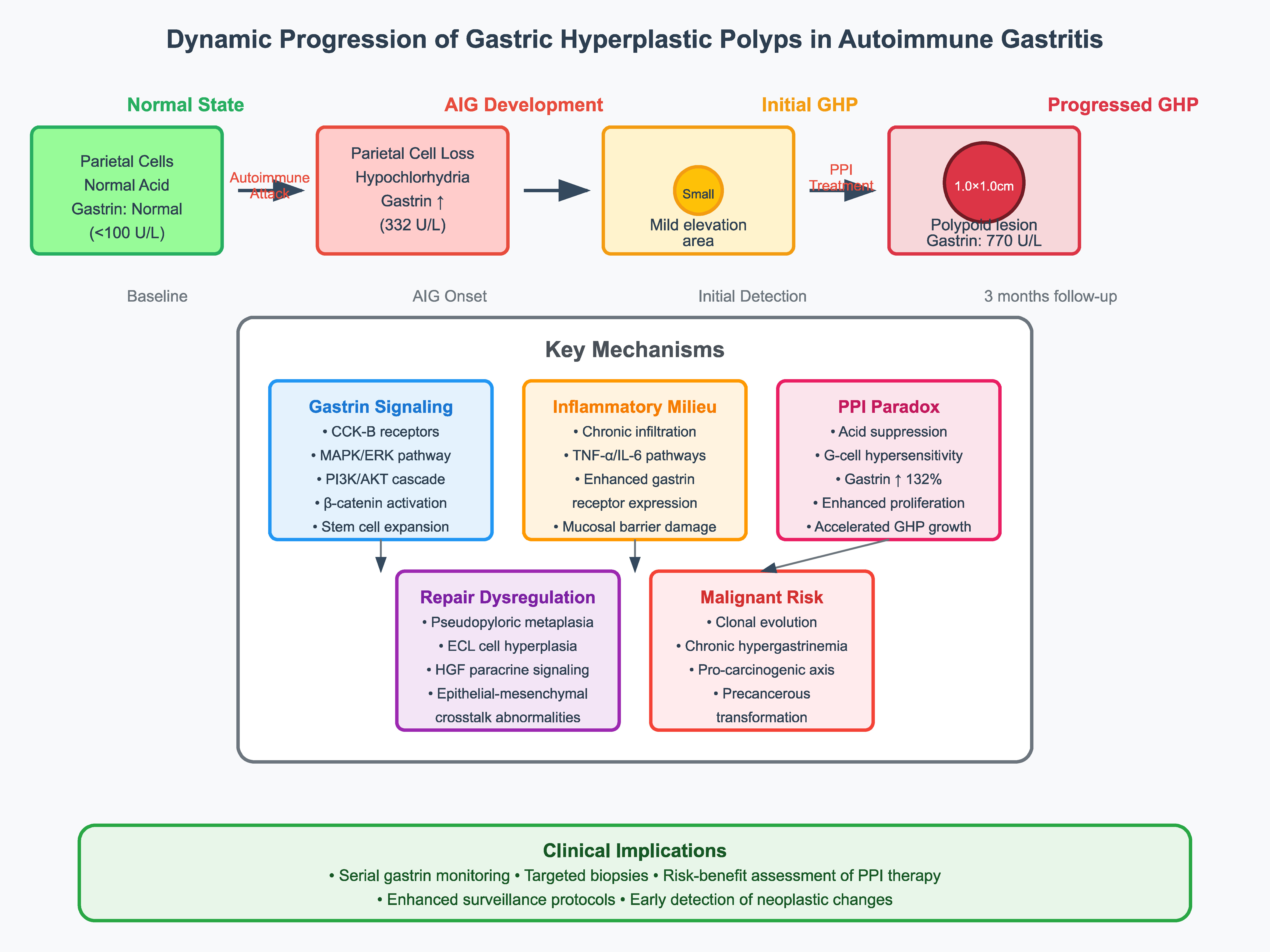
Through longitudinal clinical follow-up of a 75-year-old male with autoimmune gastritis (AIG) complicated by gastric hyperplastic polyps (GHPs), this study systematically investigated the critical regulatory role of gastrin signaling networks in gastric mucosal remodeling. The case demonstrated that within the established pathological cascade of AIG (from parietal cell destruction to gastric acid deficiency and hypergastrinemia), GHPs exhibited short-term volumetric growth. This observation challenges traditional views about the indolent nature of hyperplastic polyps. Our findings highlight the dual role of proton-pump inhibitor (PPI) therapy in AIG management while alleviating mucosal inflammation, it paradoxically induces G-cell hypersensitivity that elevates gastrin levels beyond biological thresholds (332 increasing to 770 U/L), creating a critical driver for GHP progression. This case redefines clinical management paradigms for AIG complications and offers translational insights for gastrointestinal tumor surveillance.
Keywords:autoimmune gastritis, gastric hyperplastic polyp, proton-pump inhibitor
Introduction
Autoimmune gastritis (AIG) has garnered significant research interest among chronic gastric disorders due to its distinct pathophysiological mechanisms. AIG initiates with autoimmune-mediated destruction of parietal cells, progressing through a characteristic sequence of hypochlorhydria, compensatory hypergastrinemia, and glandular atrophy - ultimately predisposing patients to pernicious anemia and gastric carcinogenesis [1,2] . GHPs, commonly associated with AIG, show higher prevalence in these patients compared to the general cohort [3] . These polyps typically develop in the context of chronic gastritis, Helicobacter pylori (H. pylori) infection, and mucosal repair processes, histologically characterized by foveolar epithelial hyperplasia, stromal edema, and inflammatory infiltration [4] . Notably, though GHPs generally exhibit indolent growth patterns, certain cases - particularly those complicated by AIG - may demonstrate accelerated progression as documented in our clinical observation.
This study reported a rapid progression of GHPs during the course of AIG in a 75-year-old male patient, which systematically reveals a central regulatory role of the gastrin signaling pathway by integrating endoscopic imaging, histopathology, and laboratory findings, providing a new perspective to understand the dynamic evolution of this type of lesion.
Case
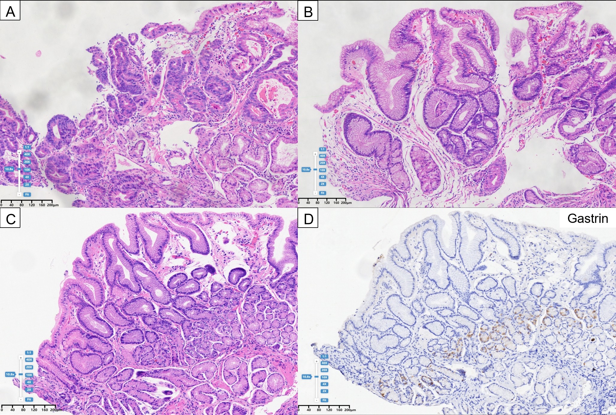
A 75-year-old male patient presented to our hospital with upper abdominal discomfort in 2023. He had no history of other systemic diseases and H. pylori eradication treatment. Endoscopic imaging evaluation revealed extensive mucosal atrophy in the greater curvature side of the gastric body (Figure 1A). A 1.2cm×1.0 cm elevated depressed lesion was detected in the upper part of the lesser curvature of the gastric body (Figure 1B). Biopsy results suggested it is a well-differentiated adenocarcinoma (Figure 2A). There was also extensive atrophy on the lesser curvature side of the gastric body with a mildly elevated area of the mucous membranes in the lower part (Figure 1C, D),designated by directional annotation markers. Histological examination showed significant proliferation in the gastric foveolar (Figure 2B). Marked atrophy was observed in the gastric angle, and the gastric antrum area was also atrophic (Figure 1E, F). Atrophic changes were present in the gastric antrum-sinus region (Figure 1E, F). Histology of the gastric antrum did not show significant atrophic intestinal epithelial chemotaxis features, but prolongation of the gastric foveolar was noted (Figure 2C). Gastrin staining indicated abnormal proliferation of gastric antrum G cells (Figure 2D). Serological tests revealed a serum gastrin level of 332 U/L, suggesting hypergastrinogenemia. Blood counts and vitamin B12 levels were normal.
Figure 1 The first endoscopic imaging evaluation A. Gastric body mucosa along the greater curvature demonstrating mucosal atrophy. B. Depressed lesion with raised margins in the superior lesser curvature of gastric body. C-D. Mild mucosal elevation in the inferior gastric body; E-F. Atrophic changes observed at the gastric angle and antrum)
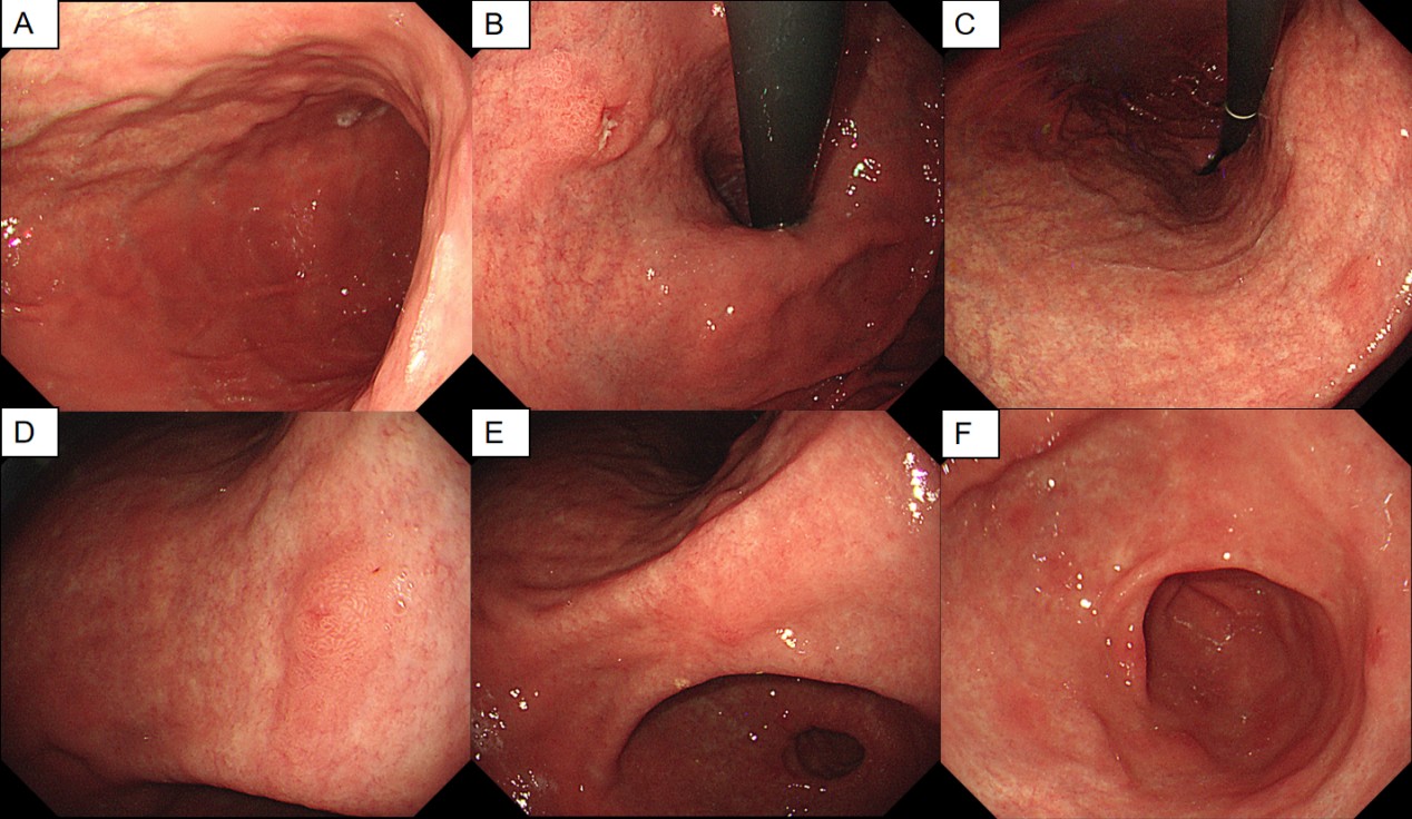
Figure 2 Pathological section A. Hematoxylin-eosin (HE) staining of superior lesser curvature lesion demonstrating well-differentiated adenocarcinoma. B. HE-stained section from inferior lesser curvature showing foveolar hyperplasia. C. Antral HE specimen revealing foveolar hyperplasia without atrophy or intestinal metaplasia. D. Gastrin staining indicated abnormal proliferation of gastric antrum G cells.
Then, the patient underwent endoscopic submucosal dissection (ESD) and 3-month postoperative proton pump inhibitor (PPI) treatment in other hospitals (esomeprazole 20mg twice daily from September 4 to November 20 in 2023), and the treatment process was uneventful.
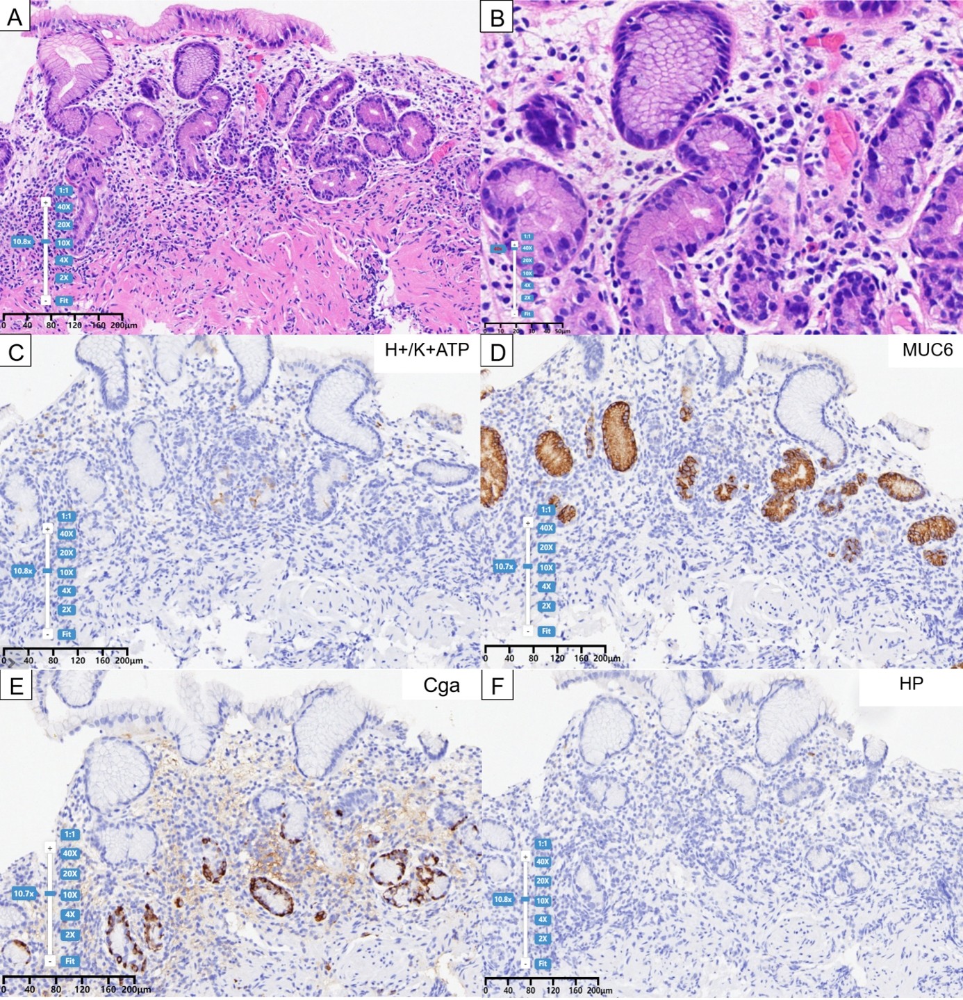
Last year the patient returned to our hospital for a gastroscopy review. Endoscopic imaging evaluation showed that the tumor in the upper part of the gastric body had been completely resected. However, extensive mucosal atrophy still persisted in the gastric body. A close - up view of the gastric body mucosa revealed the presence of Remnant oxyntic mucosa (Rom) (Figure 3A-C). Notably, the previously elevated area in the lower part of the gastric body has progressed to a 1.0 × 1.0 cm polypoid lesion (Figure 3D, E),designated by directional annotation markers. Extensive atrophy of the gastric antrum mucosa was still visible (Figure 3F). Histopathological examination of the gastric body mucosa showed mural cell destruction accompanied by deep lymphocytic infiltration and pseudo-pyloric glandular metaplasia (Figure 4A,4B). H+/K+ATPase staining revealed mural cell destruction (Figure 4C), MUC6 staining demonstrated focal positivity ( Figure 4D). Chromogranin A (CgA) staining indicated abnormal proliferation of neuroendocrine cells (Figure 4E). H. pylori staining revealed absence of H. pylori (Figure 4F).
Figure 3 The second endoscopic imaging evaluation A. Mucosal atrophy in gastric body greater curvature. B. the gastric body mucosa revealed the presence of Remnant oxyntic mucosa. C. Mucosal atrophy of superior lesser curvature. D-E. Hyperplastic polyps in inferior lesser curvature; F. Antral mucosal atrophy.
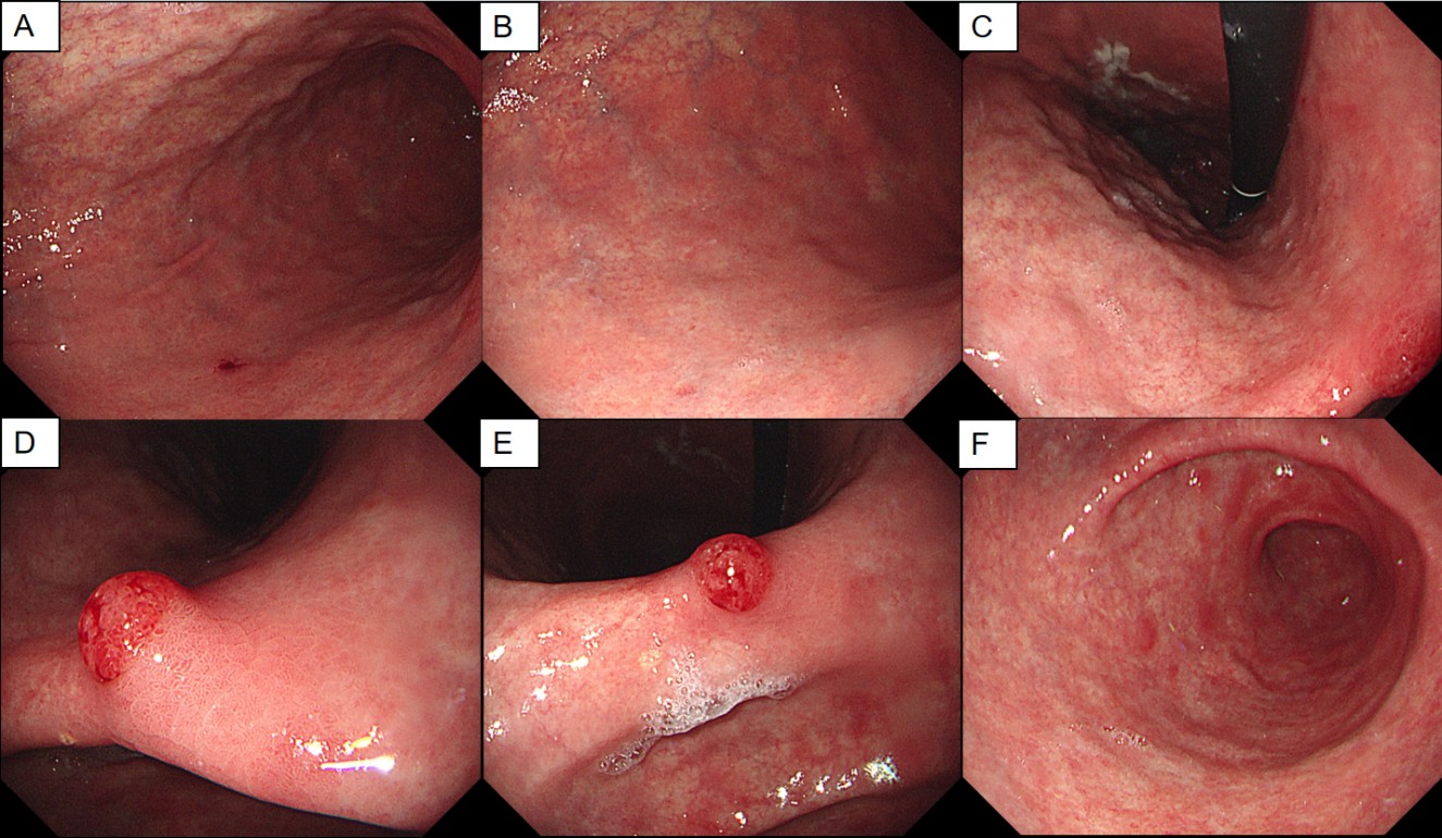
Figure 4 Pathological section A. HE-stained section from upper gastric body greater curvature showing dense lymphocytic infiltration, parietal cell loss, and pseudopyloric metaplasia. B. High-definition magnified view of Image A.C. H+/K+-ATPase staining revealed that confirming parietal cell depletion. D. MUC6 staining demonstrated focal positivity. E. CgA staining exhibited neuroendocrine cell hyperplasia. F. H. pylori staining revealed absence of H. pylori.
Serological tests revealed a positive outcome for the antiparietal cell anti-body and negative results for anti-intrinsic factor antibody. Gastrin levels spiked to 770 U/L, a 132% increase from the baseline (332 U/L vs. 770 U/L), which far exceeded the range of fluctuations commonly seen in patients with AIG (usually <500 U/L), suggesting the presence of additional stimuli such as PPIs.
In summary, the patient was diagnosed with AIG through the following features: (i) Medical history: no history of H. pylori infection. (ii) Serology: PCA positive. (iii) Histopathology: histopathological examination of the gastric body mucosa showed mural cell destruction accompanied by deep lymphocytic infiltration and pseudo-pyloric glandular metaplasia with gastric neuroendocrine cell hyperplasia. Combined with the characteristic endoscopic and pathological changes, the patient was diagnosed with AIG combined with GHPs. This case is unique because the polyps in the lower region of the gastric body shows a short- term volume doubling phenomenon, which reveals a dynamic progression of AIG related mucosal lesions.
Discussion
AIG is an organ-specific autoimmune disorder characterized by chronic inflammation of the gastric corpus mucosa and progressive glandular atrophy. Gastrin, a polypeptide hormone produced by antral G cells, plays a key physiological role in regulating gastric acid secretion and mucosal proliferation. In AIG, markedly elevated gastrin levels are closely linked to the development and progression of gastric hyperplastic polyps (GHPs) through the following mechanisms: dual activation of gastrin signaling pathways, inflammatory microenvironment and impaired mucosal repair, Paradoxical effects of PPI therapy and re-evaluating malignant transformation risks.
As a central regulator of gastric mucosal homeostasis, gastrin activates both MAPK/ERK and PI3K/AKT signaling cascades through CCK-B receptors, promoting epithelial cell proliferation and suppressing apoptosis [5] . In AIG, parietal cell loss leads to achlorhydria, which removes the normal negative feedback regulation of G cells and results in sustained hypergastrinemia demonstrated by 132% elevated levels in this case [6] . This pathological state induces abnormal β-catenin pathway activation, driving excessive expansion of gastric stem cell populations - a critical cellular basis for GHP formation [7] .
Chronic inflammatory infiltration not only directly damages the mucosal barrier but also enhances gastrin receptor expression via TNF-α/IL-6-mediated pathways, creating a pro-proliferative feedback loop [8-9] . The co-occurrence of pseudopyloric metaplasia and neuroendocrine cell hyperplasia observed in this case highlights dysregulated repair mechanisms: metaplastic epithelium loses normal secretory functions, while hyperplastic ECL cells exacerbate epithelial-mesenchymal crosstalk abnormalities through paracrine HGF secretion [10] .
Pharmacologic interventions may worsen mucosal proliferation imbalances. While PPIs can alleviate AIG-related mucosal injury symptoms, their potent acid suppression exacerbates hypergastrinemia [11] . The post-treatment exponential gastrin elevation in this case suggests PPIs may potentiate proliferative signaling through G-cell hypersensitivity [12] . This observation warrants cautious risk-benefit assessment of PPI use during rapid GHP progression.
Although traditionally considered benign, our findings suggest potential clonal evolution within GHPs under chronic hypergastrinemia. Therefore, AIG-associated GHP surveillance should integrate serial gastrin monitoring and targeted biopsies to detect early neoplastic changes.
Conclusion
This case delineates the dynamic progression of GHPs in AIG, highlighting that under PPI therapy,the central role of dysregulated gastrin signaling. The triad of persistent hypergastrinemia, chronic inflammatory milieu, and aberrant mucosal repair forms a "pro-carcinogenic axis" that facilitates the transition from benign hyperplasia to precancerous transformation. Future investigations should prioritize exploring gastrin receptor antagonists as therapeutic targets and elucidating epigenetic regulatory mechanisms underlying polyp malignant transformation, thereby establishing a scientific foundation for precision management of AIG-related complications.
Declarations
Author contributions
Yiming Song designed this study. Jianing Yan wrote the manuscript. The final manuscript has been approved by all authors.
Acknowledgements
We thank the Home for Researchers editorial team (www.home-for-researchers.com) for the language editing service. This study was supported by grants from the the Medical and Health Research Project of Zhejiang Province (No. 2024KY1515).
Funding information
This study was supported by grants from the the Medical and Health Research Project of Zhejiang Province (No. 2024KY1515).
Ethics approval and consent to participate
Not applicable.
Competing Interests
The authors declare that they have no existing or potential commercial or financial relationships that could create a conflict of interest at the time of conducting this study.
Data Availability
All data needed to evaluate the conclusions in the paper are present in the paper or the Supplementary Materials. Additional data related to this paper may be requested from the authors.
References
[1] Toh B H. Diagnosis and classification of autoimmune gastritis[J]. Autoimmunity reviews, 2014, 13(4-5): 459-462.
[2] Neumann W L, Coss E, Rugge M, et al. Autoimmune atrophic gastritis—pathogenesis, pathology and management[J]. Nature reviews Gastroenterology & hepatology, 2013, 10(9): 529-541.
[3] Carmack S W, Genta R M, Schuler C M, et al. The current spectrum of gastric polyps: a 1-year national study of over 120,000 patients[J]. Official journal of the American College of Gastroenterology| ACG, 2009, 104(6): 1524-1532.
[4] Dirschmid K, Platz-Baudin C, Stolte M. Why is the hyperplastic polyp a marker for the precancerous condition of the gastric mucosa?[J]. Virchows Archiv, 2006, 448: 80-84.
[5] Dockray G, Dimaline R, Varro A. Gastrin: old hormone, new functions[J]. Pflügers Archiv, 2005, 449: 344-355.
[6] Torbenson M, Abraham S C, Boitnott J, et al. Autoimmune gastritis: distinct histological and immunohistochemical findings before complete loss of oxyntic glands[J]. Modern pathology, 2002, 15(2): 102-109.
[7] Vannella L, Lahner E, Osborn J, et al. Risk factors for progression to gastric neoplastic lesions in patients with atrophic gastritis[J]. Alimentary pharmacology & therapeutics, 2010, 31(9): 1042-1050.
[8] Park D Y, Lauwers G Y. Gastric polyps: classification and management[J]. Archives of pathology & laboratory medicine, 2008, 132(4): 633-640;
[9] Shah S C, Piazuelo M B, Kuipers E J, et al. AGA clinical practice update on the diagnosis and management of atrophic gastritis: expert review[J]. Gastroenterology, 2021, 161(4): 1325-1332. e7.
[10] Kanayama M, Takahara T, Yata Y, et al. Hepatocyte growth factor promotes colonic epithelial regeneration via Akt signaling[J]. American Journal of Physiology-Gastrointestinal and Liver Physiology, 2007, 293(1): G230-G239.
[11] Lundell L, Vieth M, Gibson F, et al. Systematic review: the effects of long‐term proton pump inhibitor use on serum gastrin levels and gastric histology[J]. Alimentary pharmacology & therapeutics, 2015, 42(6): 649-663.
[12] Toh BH. Pathophysiology of autoimmune gastritis. Semin Immunopathol. 2014;36(5):551-562.
 Figures
 References
 Peer
 Information
Figure 1 The first endoscopic imaging evaluation A. Gastric body mucosa along the greater curvature demonstrating mucosal atrophy. B. Depressed lesion with raised margins in the superior lesser curvature of gastric body. C-D. Mild mucosal elevation in the inferior gastric body; E-F. Atrophic changes observed at the gastric angle and antrum)
Figure 2 Pathological section A. Hematoxylin-eosin (HE) staining of superior lesser curvature lesion demonstrating well-differentiated adenocarcinoma. B. HE-stained section from inferior lesser curvature showing foveolar hyperplasia. C. Antral HE specimen revealing foveolar hyperplasia without atrophy or intestinal metaplasia. D. Gastrin staining indicated abnormal proliferation of gastric antrum G cells.
Figure 3 The second endoscopic imaging evaluation A. Mucosal atrophy in gastric body greater curvature. B. the gastric body mucosa revealed the presence of Remnant oxyntic mucosa. C. Mucosal atrophy of superior lesser curvature. D-E. Hyperplastic polyps in inferior lesser curvature; F. Antral mucosal atrophy.
Figure 4 Pathological section A. HE-stained section from upper gastric body greater curvature showing dense lymphocytic infiltration, parietal cell loss, and pseudopyloric metaplasia. B. High-definition magnified view of Image A.C. H+/K+-ATPase staining revealed that confirming parietal cell depletion. D. MUC6 staining demonstrated focal positivity. E. CgA staining exhibited neuroendocrine cell hyperplasia. F. H. pylori staining revealed absence of H. pylori.
[1] Toh B H. Diagnosis and classification of autoimmune gastritis[J]. Autoimmunity reviews, 2014, 13(4-5): 459-462.
[2] Neumann W L, Coss E, Rugge M, et al. Autoimmune atrophic gastritis—pathogenesis, pathology and management[J]. Nature reviews Gastroenterology & hepatology, 2013, 10(9): 529-541.
[3] Carmack S W, Genta R M, Schuler C M, et al. The current spectrum of gastric polyps: a 1-year national study of over 120,000 patients[J]. Official journal of the American College of Gastroenterology| ACG, 2009, 104(6): 1524-1532.
[4] Dirschmid K, Platz-Baudin C, Stolte M. Why is the hyperplastic polyp a marker for the precancerous condition of the gastric mucosa?[J]. Virchows Archiv, 2006, 448: 80-84.
[5] Dockray G, Dimaline R, Varro A. Gastrin: old hormone, new functions[J]. Pflügers Archiv, 2005, 449: 344-355.
[6] Torbenson M, Abraham S C, Boitnott J, et al. Autoimmune gastritis: distinct histological and immunohistochemical findings before complete loss of oxyntic glands[J]. Modern pathology, 2002, 15(2): 102-109.
[7] Vannella L, Lahner E, Osborn J, et al. Risk factors for progression to gastric neoplastic lesions in patients with atrophic gastritis[J]. Alimentary pharmacology & therapeutics, 2010, 31(9): 1042-1050.
[8] Park D Y, Lauwers G Y. Gastric polyps: classification and management[J]. Archives of pathology & laboratory medicine, 2008, 132(4): 633-640;
[9] Shah S C, Piazuelo M B, Kuipers E J, et al. AGA clinical practice update on the diagnosis and management of atrophic gastritis: expert review[J]. Gastroenterology, 2021, 161(4): 1325-1332. e7.
[10] Kanayama M, Takahara T, Yata Y, et al. Hepatocyte growth factor promotes colonic epithelial regeneration via Akt signaling[J]. American Journal of Physiology-Gastrointestinal and Liver Physiology, 2007, 293(1): G230-G239.
[11] Lundell L, Vieth M, Gibson F, et al. Systematic review: the effects of long‐term proton pump inhibitor use on serum gastrin levels and gastric histology[J]. Alimentary pharmacology & therapeutics, 2015, 42(6): 649-663.
[12] Toh BH. Pathophysiology of autoimmune gastritis. Semin Immunopathol. 2014;36(5):551-562.
Peer-review Terminology
Identity transparency: Single anonymized
Reviewer interacts with: Editor
Details
This is an open access article under the terms of the Creative Commons Attribution License(http://creativecommons.org/licenses/by/4.0/), which permits use, distribution and reproduction in any medium, provided the original work is properly cited.
Publication History
Received 2025-03-22
Accepted 2025-06-09
Published 2025-06-30