




Abstract
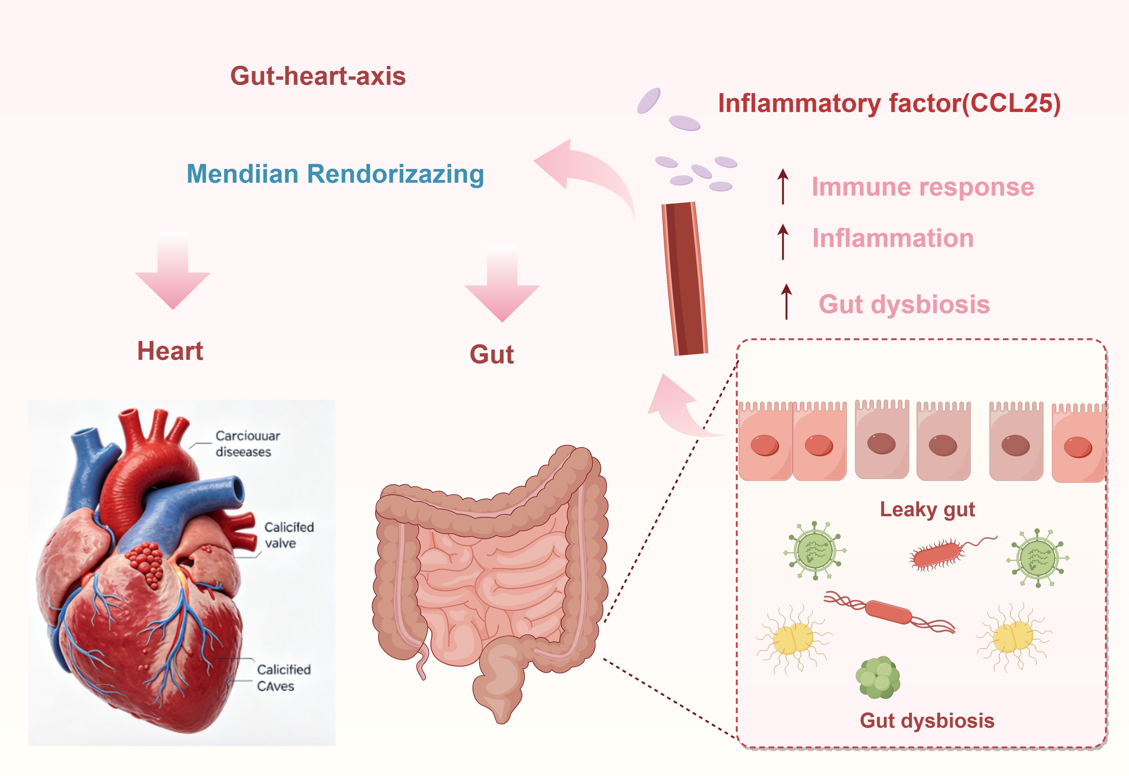
Background: Cardiovascular diseases (CVDs) arise from complex interactions between genetic predisposition and environmental exposures, manifesting as persistent inflammatory disorders. Although accumulating evidence has implicated gut microbiota in the pathogenesis of CVDs, whether this association reflects a causal relationship remains to be firmly established. Therefore, the present study sought to examine the potential causal links between gut microbiota composition, circulating inflammatory cytokines, and susceptibility to CVDs.
Methods: Summary statistics for 196 gut microbiota taxa, 91 cytokines, and 8 CVD subtypes were extracted from the largest published genome-wide association studies (GWAS) to conduct bidirectional two-sample Mendelian randomization (MR) analysis. The inverse variance weighted (IVW) method was used as the primary statistical approach. We investigated the causal associations between gut microbiota and 8 CVD subtypes, including coronary artery disease (CAD), coronary atherosclerosis (CAS), heart failure (HF), stroke, atrial fibrillation (AF), angina pectoris (AP), calcific aortic valve stenosis (CAVS), and aortic aneurysm. Multivariable MR (MVMR) analysis was further performed to explore the potential mediating role of cytokines in the causal pathways from gut microbiota to CVDs.
Results: MR analysis identified causal associations between 58 gut microbiota taxa, 46 inflammatory cytokines, and CVDs. Specifically, 34 positive and 24 negative causal effects were observed between gut microbiota and CVDs, along with 16 positive and 30 negative causal effects between cytokines and CVDs. Additionally, mediation analysis identified C-C motif chemokine 25 (CCL25) level as a key mediator in the causal relationship from gut microbiota to CVDs.
Conclusion: Our findings support a causal link between gut microbiota, inflammatory cytokines, and CVDs, with CCL25 mediating the protective effect of specific gut taxa against CAVS. These identified biomarkers offer novel insights into the pathophysiological mechanisms of CVDs and may inform the development of novel strategies for CVD prevention, diagnosis, and treatment.
Keywords: Gut microbiota; Inflammatory Cytokines; Cardiovascular diseases; Mendelian randomization
Introduction
Cardiovascular diseases (CVDs) represent a significant global health burden and are a leading cause of mortality, prompting widespread public health concern [1]. Genomics and high-throughput sequencing analysis represent contemporary methodologies for investigating the role of gut microbiota in the development of CVDs [2]. The human intestine hosts a symbiotic community of 10 to 100 trillion microbial cells that coexist with the host [3]. The gut microbiota functions as a complex ecosystem, acting as virtual endocrine organs and playing a regulatory role in the occurrence and development of CVDs [4]. One such microbiota-dependent metabolite, trimethylamine N-oxide (TMAO), has been implicated in the pathologic processes of heart failure and may serve as an early warning marker for identifying individuals at risk of disease progression [5]. The relationship between gut microbiota and heart disease is elucidated through the gut-heart axis [6]. Dysbiosis of the gut microbiome has been associated with dysregulated immune activity, particularly the enhanced secretion of inflammatory cytokines implicated in the pathogenesis of cardiovascular diseases [7]. Despite the acknowledged role of inflammation in CVD progression, studies examining the interplay between gut microbiota and cytokine profiles among CVD patients remain scarce.
Changes in gut microbiota abundance are implicated in the onset of inflammation, a key factor in the pathophysiology of CVDs [8]. Epidemiological research has identified key interleukins as playing a role in the pathogenesis of CVDs [9]. It appears that both gut microbiota and cytokines can influence the progression of CVDs. It is hypothesized that cytokines may serve as mediators in the pathway linking gut microbiota to CVDs.
Randomized controlled trials (RCTs) have traditionally been utilized to establish causal relationships between gut microbiota or cytokines and CVDs. Nevertheless, ethical considerations and financial constraints restrict the widespread implementation of RCTs in clinical settings. Mendelian randomization (MR) analysis has emerged as a valuable tool for addressing issues of causality in epidemiological data by utilizing genetic variants as instrumental variables [10]. The advancement of Genome-wide Association Studies (GWASs) and increased accessibility to GWAS data have significantly enhanced the methodology of MR analysis in recent years. While numerous studies have identified protective and pathogenic relationships between gut microbiota and cardiovascular diseases (CVDs), the potential role of inflammatory cytokines as mediators in these relationships remains unexplored [11].
This study aimed to systematically evaluate the causal links between gut microbiota composition, inflammatory cytokines, and 8 CVD subtypes via bidirectional two-sample MR and MVMR analysis, with a secondary objective to explore the mediating role of inflammatory cytokines in the gut microbiota-CVD pathways.
Methods
Study design
This study employed multiple single-nucleotide polymorphisms (SNPs) as instrumental variables
under three core assumptions to evaluate the causal effects of gut microbiota and inflammatory cytokines on eight
cardiovascular diseases: (i) the relevance assumption, which dictates that IVs must be highly associated with the
exposure; (ii) the independence assumption, which stipulates that IVs are not influenced by confounding variables; and
(iii) the exclusion restriction assumption, which posits that IVs impact the outcome solely through the exposure and
not through alternative pathways [12]. In our study, we employed bidirectional two-sample MR
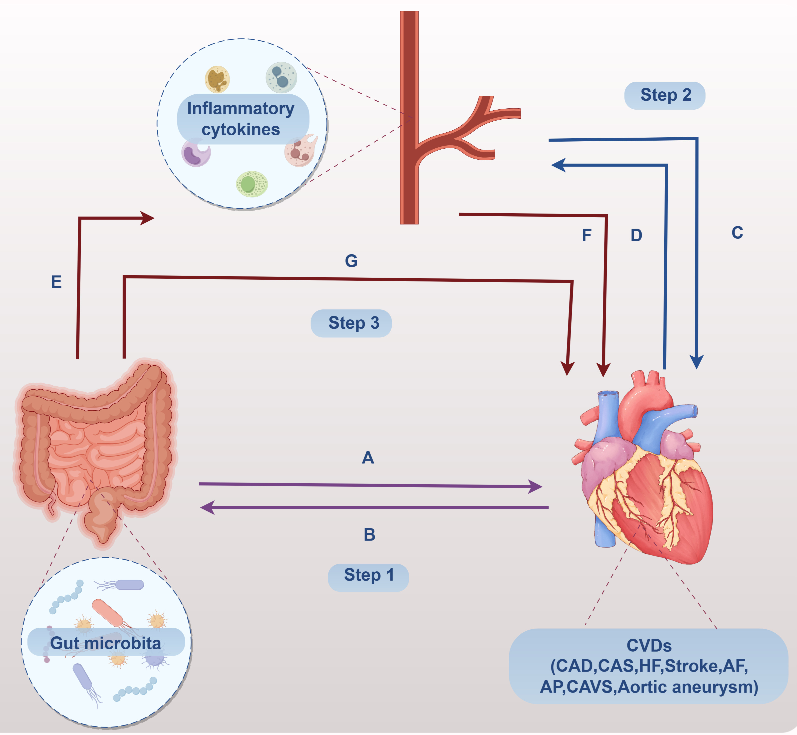
analysis and MVMR analysis, following a structured approach illustrated in Figure 1. Specifically,
Step 1 involved evaluating the bidirectional causal relationships between 196 gut microbiota and 8 cardiovascular
diseases (CVDs); Step 2 entailed identifying bidirectional causality between 91 inflammatory cytokines and 8 CVDs;
Step 3 focused on screening the 91 inflammatory cytokines as potential mediators in the pathways linking gut
microbiota to CVDs. To estimate and quantify mediator effects, we utilized two-step MR and MVMR methodologies. Of
note, bidirectional MR analysis used exactly the same IV selection criteria, statistical methods (IVW as the primary
approach), and significance threshold (P < 0.05) as the forward causal analysis to ensure the comparability of
forward and reverse results.
Figure 1. Overview of the study design for the bidirectional two-sample Mendelian randomization (MR) and mediation analysis. The study included three core steps: Step 1, bidirectional two-sample MR analysis to assess the causal relationship between 196 gut microbiota taxa and 8 CVD subtypes; Step 2, bidirectional two-sample MR analysis to assess the causal relationship between 91 circulating inflammatory cytokines and 8 CVD subtypes; Step 3, two-step mediation MR and multivariate MR (MVMR) analysis to identify the mediating role of inflammatory cytokines in the causal pathway from gut microbiota to CVDs.
Data sources
This study utilized summary statistics obtained from publicly available data from Genome-Wide
Association Studies (GWAS) conducted on populations of European ancestry. Detailed information regarding ethical
approval and informed consent can be provided in the relevant publications.
Genetic data for gut microbiota were obtained from previously published GWAS summary statistics curated by the MiBioGen consortium, which analyzed genome-wide genotypes and 16S fecal microbiome data from 18,340 individuals across 24 cohorts to assess host genetic influences on microbial composition [13]. The dataset encompassed 196 known bacterial taxa, spanning 9 phyla, 16 classes, 20 orders, 32 families, and 119 genera. Summary-level genetic data for inflammatory cytokines were derived from a separate study involving 8,293 participants, which profiled 91 cytokines and growth factors [14]. The data sources for CAD, CAS, HF, stroke, AF, AP, CAVS and aortic aneurysm used in this study are shown in Table 1.
Table 1. Summary of the GWAS data used in the MR analysis.
| Data Source | GWAS ID | Trait | Sample size | Year | Population |
|---|---|---|---|---|---|
| IEU Open GWAS | ebi-a-GCST005195 | Coronary artery disease (CAD) | 122,733 | 2017 | European |
| ebi-a-GCST009541 | Heart failure (HF) | 977,323 | 2020 | European | |
| ebi-a-GCST90018793 | Angina pectoris (AP) | 470,931 | 2021 | European | |
| ebi-a-GCST90018783 | Aortic aneurysm | 479,194 | 2021 | European | |
| FINNGEN | finn-b-I9_CORATHER | Coronary atherosclerosis (CAS) | 211,203 | 2021 | European |
| finn-b-C_STROKE | Stroke | 180,862 | 2021 | European | |
| finn-b-I9_AF | Atrial fibrillation (AF) | 138,994 | 2021 | European | |
| finngen_R9_I9_CAVS_OPERATED | Calcific aortic valve stenosis (CAVS) | 377,277 | 2019 | European |
Instrumental variables selection
Instrumental variable selection proceeded in two steps. First, SNPs
significantly associated with gut microbiota were identified using a genome-wide significance threshold of P <
1×10⁻⁵. For inflammatory cytokines, a threshold of P < 5×10⁻⁶ was applied to retain a sufficient number of valid
IVs, given the smaller sample size of the cytokine GWAS; this stricter threshold also mitigates the risk of weak
instrument bias [15]. Second, to ensure SNP independence, linkage disequilibrium clumping was
performed with an r² threshold of 0.001 within a 1000 kb window, using the 1000 Genomes Project European population
as the reference panel [16]. To ensure that the effects of SNPs on exposure and outcome were
influenced by the same allele, we performed data harmonization; palindromic SNPs with ambiguous allele frequencies were
excluded from the analysis to avoid bias from mismatched effect directions [17]. Third, the genetic
instrument's effectiveness was assessed through the F-statistic, and SNPs with an F-statistic greater than 10 were
selected to prevent bias from a weak instrument [18]. The F-statistic for each SNP was calculated
as F = R²(N−2)/(1−R²), where R² = β²/(β² + se² × N) [19]; β denotes the effect size and se the
standard error.
MR analysis
Bidirectional causality analysis. We conducted two-sample MR analysis to estimate the bidirectional causal
effects between gut microbiota abundance, inflammatory cytokines and CVDs, respectively step 1 and step 2 in
Figure 1. We used the inverse variance weighted (IVW) method as a main method for MR analysis
because it allows for robust causal estimation despite heterogeneity [20]. A pleiotropy-robust MR
analysis was conducted using four MR methods (weighted median, weighted mode, MR-Egger, and MR-PRESSO) to investigate
whether pleiotropy could contribute to MR estimation bias [21]. Mendelian randomization estimates
are presented as odds ratios with 95% confidence intervals. A causal association was considered significant when the
inverse-variance weighted method yielded P < 0.05 and the direction of effect was consistent with that obtained from
MR-Egger regression.
Mediation analysis. A two-step MR design was used to test the causal mediation effect of inflammatory cytokines (mediator) on the relationship between the gut microbiota (exposure) and CVDs (outcome), as step 3 shown in Figure 1. A two-step approach has fewer biases than a multivariable approach [22]. In the first step of two-step MR, we perform two-sample MR to estimate the causal effect between exposure and mediator, mediator and outcome, exposure and outcome separately. In the second step, we conducted a MVMR between exposure and mediator to outcome, in order to explore whether inflammatory cytokines are confounding factors affecting the causal relationship between gut microbiota and CVDs. We evaluated mediation effects using the product of coefficients method (βE × βF) with 95% CIs estimated via the delta method, which directly assessed the significance of the mediation effect that gut microbiota on CVDs through inflammatory cytokines. The total effect represents βG + βE × βF [23]. Mediation effect percentage is measured by the mediated proportion = (βE × βF)/(βG) [24].
Sensitivity analysis
Sensitivity analysis was conducted to assess the robustness of the causal
associations. In the results, we used the Cochran's Q test [25] to assess heterogeneity between
SNPs. Scatter plots were used to visualize the SNP-to-exposure and SNP-to-outcome effects, at the same time,
leave-one-out analyses were performed to determine whether the association was driven by a single SNP
[26]. To assess horizontal pleiotropy, we employed the MR-Egger regression intercept and the
MR-PRESSO global test [27]; outlier SNPs identified via MR-PRESSO were removed to correct for
horizontal pleiotropy when necessary. Sensitivity analyses were also performed for all mediation pathways to validate
the robustness of the mediation effects, with no significant heterogeneity or horizontal pleiotropy detected for the
significant pathways [28].
All analyses were conducted using the TwoSampleMR (version 0.5.8) package and MRPRESSO (version 1.0) packages in R software (Version 4.3.2).
Results
Instrumental variable selection
First, we screened 2559 SNPs (Supplementary
Table 1) from 196 bacterial taxa as IVs with a significance threshold of P < 1 × 10⁻⁵. To obtain more
comprehensive information, we performed LD-clumping (r² < 0.001, distance = 10,000 kb)
[29], harmonizing and testing pleiotropy for all the IVs to reduce correlations between the SNPs.
All retained IVs had an F-statistic > 10, indicating no risk of weak instrument bias. Second, we identified 1203
SNPs associated with 91 inflammatory cytokines by the threshold value of P < 5 × 10⁻⁶ (Supplementary Table 2).
Causal effects of gut microbiota and cytokines on the risk of 8 types of CVD
CAD
The results for the primary analyses show 77 SNPs belong to six gut microbial taxa (including 3
families, 2 genus, and 1 order) were associated with CAD (Supplementary Table 3,
Figure 2). The detailed information of the 77 SNPs is shown in Supplementary Table 4.
As presented in Figure 2, the results of the MR analyses suggested that 4 gut microbial taxa were causally associated with the risk of CAD. The family XI (OR = 1.055, 95% CI = 1.004 − 1.109, P = 0.034), family Lachnospiraceae (OR = 1.097, 95% CI = 1.019 − 1.181, P = 0.014), family Veillonellaceae (OR = 1.065, 95% CI = 1.011 − 1.122, P = 0.019), genus Oxalobacter (OR = 1.062, 95% CI = 1.019 − 1.106, P = 0.004) increased risk for developing CAD. At the same time, genetic prediction of two gut microbial taxa (genus Parabacteroides, order Lactobacillales) was associated with a significantly lower risk of CAD. The genus Parabacteroides (OR = 0.866, 95% CI = 0.784 − 0.956, P = 0.005), order Lactobacillales (OR = 0.866, 95% CI = 0.784 − 0.956, P = 0.005) decreased the risk of CAD.
As shown in Figure 3, Supplementary Table 4, the MR analysis results indicated that 9 cytokines may increase the incidence of CAD. The T-cell surface glycoprotein CD5 levels (OR = 1.161, 95% CI = 1.032 − 1.308, P = 0.013), Hepatocyte growth factor levels (OR = 1.121, 95% CI = 1.007 − 1.247, P = 0.036) significantly increased the risk of CAD. The Interleukin-5 levels (OR = 0.889, 95% CI = 0.813 − 0.973, P = 0.011), Interleukin-8 levels (OR = 0.942, 95% CI = 0.888 − 1.000, P = 0.049), Neurturin levels (OR = 0.907, 95% CI = 0.835–0.985, P = 0.021) decreased the risk of CAD.
CAS
A total of 7 gut microbiota (including 1 class, 1 family, 1 genus, 2 orders and 2 phyla) were
causally related to CAS (Supplementary Table 3, Figure 2). Details of
related 76 SNPs are shown in Supplementary Table 4.
Figure 2 shows the genus Turicibacter (OR = 1.906, 95% CI = 1.014 − 1.184, P = 0.022) suggested to increase the risk of CAS by the IVW method. Six gut microbial taxa (class Gammaproteobacteria, family Bifidobacteriaceae, order Bifidobacteriales, order Burkholderiales, phylum Actinobacteria, phylum Firmicutes) had a decreased risk of CAS. The class Gammaproteobacteria (OR = 0.829, 95% CI = 0.727 − 0.945, P = 0.005) and phylum Firmicutes (OR = 0.876, 95% CI = 0.786 − 0.976, P = 0.017) significantly decreased the risk of CAS.
Figure 3 shows that C-C motif chemokine 4 levels, Fibroblast growth factor 5 levels and Interleukin-22 receptor subunit alpha-1 levels seemed to be risk factors to CAS. The Interleukin-22 receptor subunit alpha-1 levels significantly increased the incidence of CAS. The Leukemia inhibitory factor receptor levels was a protective factor for CAS.
HF
A total of 9 gut microbiota (including 2 class, 4 genus and 3 orders, 2 phyla) were causally
associated with HF (Supplementary Table 3, Figure 2). Details of
related 91 SNPs are shown in Supplementary Table 4.
As Figure 2 shows, five gut microbial taxa (class Negativicutes, genus Eubacterium eligens group, genus Eubacterium oxidoreducens group, genus Flavonifractor, order Selenomonadales) seemed to be risk factors to HF. The genus Eubacterium eligens group (OR = 1.126, 95% CI = 1.017 − 1.247, P = 0.023) and genus Flavonifractor (OR = 1.144, 95% CI = 1.031 − 1.270, P = 0.012) considerably elevated risk for HF. Four gut microbial taxa (class Bacteroidia, genus Anaerostipes, order Bacillales and order Bacteroidales) seemed to be protective factors to HF. The genus Anaerostipes (OR = 0.899, 95% CI = 0.825 − 0.978, P = 0.014) significantly reduced the risk for HF.
In Figure 3, four cytokines (T-cell surface glycoprotein CD5 levels, Macrophage colony-stimulating factor 1 levels, Interleukin-2 levels, Tumor necrosis factor levels) were associated with an increased risk of HF. The T-cell surface glycoprotein CD5 levels (OR = 1.100, 95% CI = 1.016 − 1.191, P = 0.019) and Tumor necrosis factor levels (OR = 1.110, 95% CI = 1.029 − 1.199, P = 0.007) significantly increased the risk of HF. The Interleukin-17C levels (OR = 0.907, 95% CI = 0.832 − 0.989, P = 0.027), Leukemia inhibitory factor receptor levels (OR = 0.913, 95% CI = 0.849 − 0.981, P = 0.014) and Sulfotransferase 1A1 levels (OR = 0.950, 95% CI = 0.905 − 0.998, P = 0.041) decreased the risk of HF.
Stroke
A total of 10 gut microbiota (including 4 family, 5 genus, 1 order) had a causal relationship with
stroke (Supplementary Table 3, Figure 2). Details of related 97 SNPs
are shown in Supplementary Table 4.
As shown in Figure 2, the family Bacteroidales S24-7 group (OR = 1.109, 95% CI = 1.025 − 1.200, P = 0.010), genus Holdemanella (OR = 1.080, 95% CI = 1.014 − 1.151, P = 0.017) and genus Marvinbryantia (OR = 1.111, 95% CI = 1.002 − 1.231, P = 0.046) suggested to be risk factors for stroke. Seven gut microbial taxa (family Alcaligenaceae, family Defluviitaleaceae, family Desulfovibrionaceae, genus Eggerthella, genus Faecalibacterium, genus Ruminococcus torques group) may be protective factors for stroke. The family Desulfovibrionaceae (OR = 0.875, 95% CI = 0.783 − 0.979, P = 0.020) and genus Ruminococcus torques group (OR = 0.769, 95% CI = 0.631 − 0.936, P = 0.009) were significantly lower risk for stroke.
Figure 3 shows that C-X-C motif chemokine 11 levels (OR = 1.096, 95% CI = 1.012 − 1.187, P = 0.025), FGF5 (OR = 1.053, 95% CI = 1.022 − 1.085, P = 0.001), Interleukin-2 receptor subunit beta levels (OR = 1.123, 95% CI = 1.026 − 1.230, P = 0.012) seemed to be risk factors to stroke. The Cystatin D levels (OR = 0.963, 95% CI = 0.935 − 0.992, P = 0.014) and Stem cell factor levels (OR = 0.954, 95% CI = 0.910 − 1.000, P = 0.048) were protective factors to stroke.
AF
The results suggest a causal relationship between 9 gut microbiota (including 1 family, 5 genus, 1
order and 2 phylum) and AF (Supplementary Table 3, Figure 2). Details
of related 106 SNPs are shown in Supplementary Table 4.
As shown in Figure 2, the family Pasteurellaceae (OR = 1.107, 95% CI = 1.021 − 1.199, P = 0.013), genus Ruminiclostridium5 (OR = 1.142, 95% CI = 1.017 − 1.283, P = 0.025) and order Pasteurellales (OR = 1.107, 95% CI = 1.021 − 1.199, P = 0.013) suggest to be risk factors to AF. Six gut microbial taxa (genus Bifidobacterium, genus Eggerthella, genus Howardella, genus Ruminococcus 1, phylum Actinobacteria, phylum Firmicutes) may be protective factors for AF. The phylum Firmicutes (OR = 0.860, 95% CI = 0.750 − 0.985, P = 0.029) significantly decreased the incidence of AF.
Figure 3 shows that six inflammatory cytokines (Eotaxin levels, C-X-C motif chemokine 1 levels, C-X-C motif chemokine 11 levels, FGF5 levels, Interleukin-2 receptor subunit beta levels, TNF-beta levels) seemed to be risk factors for AF. The Interleukin-2 receptor subunit beta levels (OR = 1.131, 95% CI = 1.003 − 1.275, P = 0.045) significantly increased the risk of AF. Four inflammatory cytokines (C-C motif chemokine 25 levels, CD40L receptor levels, Leukemia inhibitory factor receptor levels, Matrix metalloproteinase-10 levels) suggested to be protective factors to AF. Among them, the Leukemia inhibitory factor receptor levels (OR = 0.871, 95% CI = 0.812 − 0.935, P < 0.001) significantly decreased the risk of AF.
AP
The IVW method revealed 52 causal associations of gut microbiota on AP, including 1 family, 1 genus, 2
orders and 1 phylum (Figure 2, Supplementary Table 3). Details of
related 52 SNPs are shown in Supplementary Table 4. The risk of AP may be increased by
three gut microbiota traits, family XI (OR = 1.056, 95% CI = 1.008 − 1.107, P = 0.022), genus Blautia (OR = 1.622,
95% CI = 1.223 − 2.151, P = 0.010), order Gastranaerophilales (OR = 1.074, 95% CI = 1.006 − 1.147, P = 0.032). The
genus Terrisporobacter (OR = 0.921, 95% CI = 0.848 − 1.000, P = 0.050), order Lactobacillales (OR = 0.913, 95% CI =
0.838 − 0.996, P = 0.040), phylum Actinobacteria (OR = 0.896, 95% CI = 0.822 − 0.975, P = 0.011) may reduce the risk
of AP.
Described in Figure 3, five inflammatory cytokines (T-cell surface glycoprotein CD5 levels, Macrophage colony-stimulating factor 1 levels, FGF5 levels, Interleukin-10 receptor subunit beta levels, Interleukin-17A levels) may increase the risk of AP (Supplementary Table 3). Among them, the Interleukin-17A levels (OR = 1.108, 95% CI = 1.012 − 1.214, P = 0.027) significantly increased the risk of AP. The Interleukin-1-alpha levels (OR = 0.943, 95% CI = 0.891 − 0.997, P = 0.040), Interleukin-6 levels (OR = 0.856, 95% CI = 0.758 − 0.967, P = 0.012), Leukemia inhibitory factor receptor levels (OR = 0.931, 95% CI = 0.875 − 0.990, P = 0.023) may decrease the risk of AP.
CAVS
Based on the IVW method, the results suggested 78 causal relationships between gut microbiota and
CAVS, including 1 family, 4 genus, 1 order and 1 phylum (Figure 2, Supplementary Table 3). Details of related 78 SNPs are shown in Supplementary Table 4. The family Desulfovibrionaceae (OR = 1.311, 95% CI = 1.088 −
1.580, P = 0.005), genus Turicibacter (OR = 1.208, 95% CI = 1.007 − 1.449, P = 0.042), order Desulfovibrionales (OR
= 1.213, 95% CI = 1.021 − 1.441, P = 0.028) seemed to be risk factors to CAVS. There were four gut microbiota (genus
Faecalibacterium, genus Ruminococcus2, genus Subdoligranulum, phylum Actinobacteria) associated with a decreased risk
of CAVS. The phylum Actinobacteria (OR = 0.841, 95% CI = 0.719 − 0.984, P = 0.031) significantly decreased risk of
CAVS.
As shown in Figure 3, four inflammatory cytokines (C-C motif chemokine 19 levels, Hepatocyte growth factor levels, Interleukin-8 levels, TNF-beta levels) seemed to increase the risk of CAVS. The Hepatocyte growth factor levels (OR = 1.202, 95% CI = 1.054 − 1.372, P = 0.006) significantly increased risk of CAVS. The C-C motif chemokine 25 levels (OR = 0.941, 95% CI = 0.901 − 0.983, P = 0.007) and Interleukin-10 levels (OR = 0.877, 95% CI = 0.770 − 0.999, P = 0.048) decreased the risk of CAVS.
Aortic aneurysm
Genetic prediction of five gut microbiota (including 1 class and 4 genus) was associated
with a causal effect of Aortic aneurysm (Figure 2, Supplementary Table
3). Details of related 68 SNPs are shown in Supplementary Table 4. The genus
Anaerofilum (OR = 1.172, 95% CI = 1.013 − 1.355, P = 0.033) and genus Ruminococcaceae UCG005 (OR = 1.434, 95% CI =
1.171 − 1.757, P < 0.001) may increase the risk of Aortic aneurysm. The class Actinobacteria (OR = 0.751, 95% CI =
0.583 − 0.967, P = 0.027), genus Prevotella 9 (OR = 0.810, 95% CI = 0.684 − 0.959, P = 0.014), genus
Ruminococcaceae NK4A214 group (OR = 0.715, 95% CI = 0.573 − 0.893, P = 0.003) may be protective factors to Aortic
aneurysm.
A total of six inflammatory cytokines were associated with Aortic aneurysm (Figure 3, Supplementary Table 4). Four inflammatory cytokines (Fractalkine levels, C-X-C motif chemokine 6 levels, FGF5 levels, Interleukin-20 receptor subunit alpha levels) were risk factors for aortic aneurysm, the Interleukin-20 receptor subunit alpha levels (OR = 1.251, 95% CI = 1.015 − 1.541, P = 0.035) significantly increased risk of aortic aneurysm. The C-C motif chemokine 25 levels (OR = 0.894, 95% CI = 0.836 − 0.956, P = 0.001) and Stem cell factor levels (OR = 0.870, 95% CI = 0.778 − 0.973, P = 0.015) had a protective causal effect on Aortic aneurysm.
Figure 2. Mendelian randomization results of the causal effects between gut microbiota taxa and 8 CVD subtypes, estimated using the inverse variance weighted (IVW) method. Results are presented as odds ratios (ORs) with 95% confidence intervals (CIs).

Figure 3. Mendelian randomization results of the causal effects between circulating inflammatory cytokines and 8 CVD subtypes, estimated using the inverse variance weighted (IVW) method.

Bi‑directional causal effects of CVDs on gut microbiota and cytokines
CAD. When assessing the bi‑directional causal effect of CAD on gut microbiota and cytokines, we found evidence
suggesting a genetic predisposition to CAD was linked to specific gut microbiota, with lower abundance in one family,
five genus and one order, and higher abundance in one family, four genus and 1 order (Supplementary Table 7). The most significant result in lower abundance was the genus
Actinomyces (OR = 0.926, 95% CI = 0.872 − 0.983, P = 0.011), and in higher abundance was the genus Gordonibacter (OR
= 1.098, 95% CI = 1.010 − 1.193, P = 0.027). CAD was associated with a lower abundance in 8 cytokines (Supplementary Table 8), the most significant result was the TNF-beta levels (OR = 0.943,
95% CI = 0.894 − 0.996, P = 0.034).
CAS. CAS has a causal relationship on nine gut microbiota (Supplementary Table 7), with lower abundance in two genus (genus Dialister (OR = 0.956, 95% CI = 0.915 − 1.000, P = 0.050), genus Family XIII UCG001 (OR = 0.927, 95% CI = 0.887 − 0.968, P = 0.001)), and with higher abundance in one class, one family, four genus and one order, the most significant result was the genus Veillonella (OR = 1.083, 95% CI = 1.030 − 1.140, P = 0.002). CAS was associated with a lower abundance in 4 cytokines (Supplementary Table 8), the most significant result was the T-cell surface glycoprotein CD5 levels (OR = 0.953, 95% CI = 0.910 − 0.999, P = 0.046).
HF. HF was associated with lower abundance in one class, two family, three genus and one order (Supplementary Table 7), the most significant result was the genus Victivallis (OR = 0.787, 95% CI = 0.635 − 0.974, P = 0.028). HF has a causal relationship with higher abundance in one family, six genus and one order, the most significant result was the genus Lachnospiraceae UCG001 (OR = 1.157, 95% CI = 1.027 − 1.303, P = 0.016). HF has a causal association with Interleukin-24 levels (OR = 1.094, 95% CI = 1.005 − 1.191, P = 0.038).
Stroke. Stroke has a causal relationship with lower abundance in two genus (genus Marvinbryantia (OR = 0.902, 95% CI = 0.816 − 0.998, P = 0.045), genus Prevotella 9 (OR = 0.888, 95% CI = 0.805 − 0.979, P = 0.017)), and with higher abundance in two class, three genus, the most significant result was the genus Lactococcus (OR = 1.230, 95% CI = 1.050 − 1.440, P = 0.01). As shown in Supplementary Table 8, there was no reverse effect between stroke and cytokines.
AF. AF was associated with lower abundance in 1 family and 7 genus (Supplementary Table 7), the most significant result was the genus Prevotella7 (OR = 0.943, 95% CI = 0.890 − 0.998, P = 0.044), and with high abundance in genus Butyricimonas (OR = 1.035, 95% CI = 1.001 − 1.069, P = 0.041) and genus Intestinibacter (OR = 1.134, 95% CI = 1.003 − 1.066, P = 0.034). As shown in Supplementary Table 8, there was no reverse effect between AF and cytokines.
AP. AP has a causal relationship with lower abundance in 7 gut microbiota (including 1 class, 1 family, 4 genus and 1 order), the most significant result was the genus Allisonella (OR = 0.891, 95% CI = 0.803 − 0.989, P = 0.030). AP was associated with higher abundance in 1 class, 1 family, 3 genus and 1 order, the most significant result was the genus Veillonella (OR = 1.097, 95% CI = 1.038 − 1.159, P = 0.001). AP was associated with a lower abundance in 8 cytokines (Supplementary Table 8), the most significant result was the T-cell surface glycoprotein CD5 levels (OR = 0.913, 95% CI = 0.879 − 0.948, P = 0.001).
CAVS. CAVS was associated with lower abundance in 7 gut microbiota (including 1 class, 1 family, 3 genus, 1 order and 1 phylum, Supplementary Table 7), the most significant result was the genus Ruminococcus gnavus group (OR = 0.947, 95% CI = 0.896 − 1.000, P = 0.005), and with high abundance in family XI (OR = 1.140, 95% CI = 1.027 − 1.264, P = 0.014). As shown in Supplementary Table 8, there was no reverse effect between CAVS and cytokines.
Aortic aneurysm. Aortic aneurysm was associated with lower abundance in 13 gut microbiota (including 2 class, 4 family, 4 genus, 2 order and 1 phylum, Supplementary Table 7), the most significant result was the genus Eubacterium nodatum group (OR = 0.906, 95% CI = 0.827 − 0.993, P = 0.035), and with high abundance in the genus Catenibacterium (OR = 1.119, 95% CI = 1.004 − 1.246, P = 0.041). Aortic aneurysm was associated with a lower abundance in 4 cytokines (Supplementary Table 8), the most significant result was the Leukemia inhibitory factor levels (OR = 0.958, 95% CI = 0.932 − 0.986, P = 0.003), and with high abundance in Interleukin-18 levels (OR = 1.031, 95% CI = 1.004 − 1.058, P = 0.024).
Sensitivity analyses
For the robustness of the estimates, we further conducted a sensitivity and
pleiotropy analysis to avoid horizontal pleiotropy, with all intercept P values > 0.05 and global test P values
> 0.05, indicating no significant horizontal pleiotropy for all identified causal associations. Cochran's Q tests
demonstrated no significant heterogeneity (P > 0.05, Supplementary Table 9).
Mediation analysis of gut microbiota, inflammatory cytokines, and CVDs
To explore the underlying potential
mechanisms involving in the occurrence and development of CVDs, we performed mediation analysis to identify the causal
pathway from gut microbiota to CVDs mediated by cytokines. Initially, the results of two-sample MR indicate that gut
microbiota has causal effects on cytokines. We identified 22 associations of gut microbiota to cytokines (CAD, 3; CAS,
3; HF, 4; Stroke, 1; AF, 5; AP, 1; CAVS, 3; Aortic Aneurysm, 2) (Supplementary Table
10). Secondly, MVMR was applied to explore the significant causal association between cytokines with CVDs after
correcting for the gut microbiota. The reliability of results was verified by sensitivity analysis, indicating that no
heterogeneity and horizontal pleiotropy existed (Supplementary Table 9).
Notably, the majority of tested pathways showed only a non-significant trend of mediation effect without statistical support, and thus were not considered as valid mediating pathways. In summary, we identified 2 mediating relationships, including 2 gut microbiota–CAVS causal pathways mediated by 1 cytokine (Table 2). The mediation analysis reveals that C-C motif chemokine 25 levels exhibit significant negative mediation effects to CAVS, respectively on genus Subdoligranulum (beta = −0.017, 95% CI [−0.035, −0.002], P = 0.034) with 9.55% proportion, and phylum Actinobacteria (beta = −0.018, 95% CI [−0.035, −0.005], P = 0.024) with 10.41% proportion.
Table 2. Mediation analysis of gut microbiota, inflammatory cytokines, and CVDs.
| Exposure | Mediator | Outcome | Total effect | Direct effect | Mediation effect (95% CI) | P value | Mediation Proportion |
|---|---|---|---|---|---|---|---|
| family Veillonellaceae | Macrophage colony-stimulating factor 1 levels | CAD | 0.063 | 0.057 | 0.006 (0, 0.016) | 0.134 | 9.55% |
| genus Parabacteroides | C-X-C motif chemokine 10 levels | CAD | −0.144 | −0.123 | −0.021 (−0.058, 0.002) | 0.189 | 14.56% |
| order Lactobacillales | Interleukin-5 levels | CAD | −0.084 | −0.076 | −0.008 (−0.024, 0.004) | 0.253 | 9.51% |
| phylum Actinobacteria | Leukemia inhibitory factor receptor levels | CAS | −0.092 | −0.079 | −0.013 (−0.033, 0) | 0.104 | 14.06% |
| genus Bifidobacterium | C-C motif chemokine 25 levels | AF | −0.132 | −0.125 | −0.007 (−0.017, 0) | 0.162 | 5.29% |
| phylum Actinobacteria | C-C motif chemokine 25 levels | AF | −0.699 | −0.690 | −0.009 (−0.023, 0) | 0.134 | 1.29% |
| phylum Actinobacteria | Leukemia inhibitory factor receptor levels | AF | −0.699 | −0.681 | −0.018 (−0.043, 0) | 0.102 | 2.57% |
| phylum Actinobacteria | Leukemia inhibitory factor receptor levels | AP | −0.110 | −0.099 | −0.011 (−0.028, 0) | 0.116 | 9.98% |
| genus Subdoligranulum | C-C motif chemokine 25 levels | CAVS | −0.178 | −0.161 | −0.017 (−0.035, −0.002) | 0.034 | 9.55% |
| phylum Actinobacteria | C-C motif chemokine 25 levels | CAVS | −0.173 | −0.155 | −0.018 (−0.035, −0.005) | 0.024 | 10.41% |
| genus Ruminococcaceae NK4A214 group | Fractalkine levels | Aortic Aneurysm | −0.390 | −0.361 | −0.029 (−0.071, −0.001) | 0.107 | 7.44% |
Discussion
Emerging evidence has established strong associations between gut microbiota-derived metabolites [30]—particularly trimethylamine-N-oxide and short-chain fatty acids—and the pathogenesis of cardiovascular diseases [31], including ischemic heart disease [32], heart failure, and hypertension [33]. Beyond metabolic pathways, the gut microbiota also exerts profound effects on host immune responses, modulating key cellular processes such as immunoregulation, apoptosis, and inflammatory signaling [34-36]. Chronic inflammation and autoimmune mechanisms have likewise been implicated in cardiovascular pathology [37, 38]. Of particular interest, recent studies have identified autoantibodies directed against C-X-C motif chemokine receptor 3, a G-protein-coupled receptor known to participate in atherosclerotic progression [39]. Nevertheless, the intricate nature of cardiovascular diseases and the diverse composition of gut microbiota present challenges in comprehensively elucidating the impact of gut microbiota on cardiovascular conditions through observational investigations. Furthermore, the potential mediating role of inflammatory factors in signaling pathways remains uncertain.
This study utilized MR analysis to assess the causal relationship between gut microbiota, inflammatory cytokines and CVDs. The investigation examined potential causal associations between 196 gut microbiota abundance variables and 8 types of CVDs (CAD, HF, AP, Aortic aneurysm, CAS, AF, CAVS), identifying a potential causal relationship involving 58 gut microbiota, 46 inflammatory cytokines, and 8 types of CVDs. Furthermore, mediation analysis results provided support for the involvement of altered inflammatory cytokines in the pathogenesis of CVDs.
Our research findings indicate a potential causal relationship between specific gut microbiota and various cardiovascular diseases, with certain gut microbiota exhibiting a consistent causal role across different types of cardiovascular diseases. Specifically, our results suggest that a high abundance of the phylum Actinobacteria may serve as a protective factor against four cardiovascular diseases (coronary artery stenosis, atrial fibrillation, aortic aneurysm, and calcific aortic valve stenosis). A clinical trial conducted in northwestern China further supported this notion, demonstrating elevated levels of Actinobacteria in the fecal samples of healthy individuals compared to those with hypertension [40]. Collinsella, a dominant taxon of the family Coriobacteriaceae within the phylum Actinobacteria [41], has been found to have elevated abundance in patients with type 2 diabetes and symptomatic carotid atherosclerotic stenosis [42, 43].
Additionally, our research indicates that FGF5 levels are a key risk factor for stroke, atrial fibrillation, angina pectoris, and aortic aneurysm. Previous studies from the 1990s have demonstrated the active role of fibroblast growth factors (FGFs) in regulating endothelial cells (EC) and smooth muscle cells (SMC) [44]. Moreover, numerous studies have demonstrated the significance of inflammatory responses in the vascular system as key contributors to atherosclerosis, with basic Fibroblast Growth Factor (bFGF) playing a role in vascular inflammation [45]. FGF5, a member of the FGF family, has been linked to vascular remodeling and hypertension in previous clinical studies, which aligns with our finding that FGF5 increases the risk of multiple CVD subtypes. Furthermore, our findings suggest that elevated levels of Macrophage colony-stimulating factor 1 may be associated with an increased risk of coronary artery disease, heart failure, and angina pectoris. Conversely, decreased levels of C-C motif chemokine 25 may be linked to a reduced risk of atrial fibrillation, calcific aortic valve stenosis, and aortic aneurysm. Prior research has demonstrated that a deficiency in M-CSF can significantly decrease the progression of atherosclerosis in mice with hyperlipidemia. Additionally, a study has demonstrated that macrophage colony-stimulating factor plays a crucial role in neointimal thickening formation following arterial injury [46]. Chemokines, in turn, contribute to disease pathogenesis by directing leukocyte migration to inflammatory sites [47, 48]. Studies utilizing serum proteomic profiling in heart failure cohorts have demonstrated an age-related increase in circulating C-C motif chemokine ligand 17 (CCL17) levels, which correlate with cardiac insufficiency [49]. Nevertheless, the impact of C-C motif chemokine 25 levels on the development of cardiovascular diseases remains poorly understood.
Furthermore, our study investigated the reverse causal effects of 8 CVD subtypes on gut microbiota and cytokine levels through bidirectional MR analysis. The findings indicated that 4 CVD subtypes exhibited bidirectional causal effects on the abundance of class Gammaproteobacteria, family Pasteurellaceae, and order Pasteurellales, while 3 CVD subtypes had bidirectional causal effects on levels of Interleukin-12 receptor subunit beta and T-cell surface glycoprotein CD5. These reverse causal findings suggest that the occurrence and progression of CVDs may further exacerbate gut microbiota dysbiosis and systemic inflammatory dysregulation, forming a vicious cycle of "microbiota dysregulation − inflammatory activation − CVD progression", which provides new insights for the long-term management of CVD patients. Additionally, we conducted further analysis to identify potential mediators of inflammatory cytokines in the pathway linking gut microbiota to cardiovascular diseases. Our research revealed that C-C motif chemokine 25 levels act as a mediator in the pathways linking the genera Subdoligranulum and phylum Actinobacteria to CAVS, with mediation effects of 9.55% and 10.41% respectively.
This study represents the initial exploration of the causal connections between gut microbiota, inflammatory cytokines, and eight CVDs utilizing a thorough and integrated MR Framework. Moreover, we established a pathway from gut microbiota to CVDs through the mediation analysis of inflammatory factors. In order to assess the validity of our findings in light of potential violations of Mendelian randomization assumptions, we conducted various sensitivity analyses using multiple Mendelian randomization approaches. Nonetheless, this study is subject to certain limitations. Firstly, the sample size for cardiovascular disease subtypes was inadequate. Secondly, the study only included populations of European descent due to limited availability of genome-wide association study data from other populations. Lastly, additional experiments and in-depth analysis of clinical patient data will be necessary to confirm the results.
Despite these advancements, our study has several limitations that warrant consideration. First, the sample sizes for CVD subtypes, particularly rare conditions such as CAVS and aortic aneurysm, were relatively small, which may reduce the statistical power to detect causal associations and increase the risk of type II errors. Second, the exclusive use of GWAS data from European populations limits the generalizability of our findings to other ethnic groups. Given the known heterogeneity in gut microbiota composition and genetic susceptibility to CVDs across populations, future studies should prioritize multi-ethnic cohorts to validate these associations. Third, while MR analysis minimizes confounding and reverse causation, it cannot fully replace experimental validation. The mechanisms linking specific bacterial taxa (e.g., Actinobacteria) to CVDs protection remain speculative, necessitating functional studies using animal models or in vitro systems to elucidate microbial metabolite production, host immune interactions, and vascular remodeling pathways.
To address these gaps, we propose the following research priorities: (1) Large-scale longitudinal studies integrating multi-omics data (metagenomics, metabolomics, and proteomics) to track dynamic microbiota-cytokine-CVD interactions; (2) Randomized controlled trials testing dietary, probiotic, or anti-cytokine interventions targeting identified mediators (e.g., CCL25 or FGF5); (3) Development of ethnic-specific microbiota biomarkers for CVD risk stratification; and (4) Mechanistic investigations into how bidirectional CVD-microbiota effects influence disease progression (e.g., through gut-barrier integrity or systemic inflammation).
Additionally, we emphasize the need for standardization in microbiota nomenclature and cytokine measurement across studies to enhance comparability. Future MR analyses should also explore sex-specific and age-stratified causal effects, given the demographic variability in CVD presentations.
Conclusion
In this study, a comprehensive Mendelian randomization analysis was conducted to investigate the relationships between gut microbiota, inflammatory cytokines, and eight types of CVDs. The analysis revealed 34 positive causal effects and 24 negative causal effects between genetic predisposition in gut microbiota and CVDs, as well as 16 protective factors and 30 risk factors between cytokines and CVDs. We further identified two significant mediating pathways, in which CCL25 mediated the protective effect of Subdoligranulum and Actinobacteria against CAVS. The gut microbiota taxa and inflammatory cytokines identified in this investigation may serve as novel biomarkers for CVD risk stratification and management, as well as enhance our understanding of the pathophysiological mechanisms underlying CVDs. In conclusion, our findings provide novel genetic evidence for the gut-heart axis, while the outlined limitations highlight the necessity for translational research to bridge these genetic associations with clinical applications.
Abbreviations
AF, Atrial Fibrillation; AP, Angina Pectoris; CCL17, C-C Motif Chemokine Ligand 17; CCL25, C-C Motif Chemokine 25; CAD, Coronary Artery Disease; CAS, Coronary Atherosclerosis; CAVS, Calcific Aortic Valve Stenosis; CI, Confidence Interval; CVDs, Cardiovascular Diseases; EC, Endothelial Cells; FGF5, Fibroblast Growth Factor 5; GWAS, Genome-Wide Association Studies; HF, Heart Failure; IV, Instrumental Variable; IVW, Inverse Variance Weighted; LD, Linkage Disequilibrium; M-CSF, Macrophage Colony-Stimulating Factor 1; MR, Mendelian Randomization; MVMR, Multivariable Mendelian Randomization; OR, Odds Ratio; RCT, Randomized Controlled Trial; SMC, Smooth Muscle Cells; SNP, Single-Nucleotide Polymorphism; TMAO, Trimethylamine N-Oxide.
Supplementary Materials
Declarations
Author Contributions
Xuekun Hou: Conceptualization, Methodology, Formal Analysis, Writing – Original Draft Preparation. Yunxiao Meng: Methodology, Formal Analysis, Writing – Original Draft Preparation. Yuxuan Zhang: Formal Analysis, Data Curation. Xianghan Zheng: Formal Analysis, Data Curation. Hongqi Guo: Validation, Visualization. Yichen Zhang: Validation, Visualization. Hongmei Yuan: Project Administration, Writing – Review & Editing. Zefei Chu: Conceptualization, Writing – Review & Editing, Supervision, Funding Acquisition. All authors read and approved the final manuscript.
Acknowledgements
Not Applicable.
Funding Information
The project was financially supported by Medical Science Research Project of Hebei Provincial Health Commission (20251061/20251060); Scientific Research Project for Institutions of Higher Education, Department of Education of Hebei Province (QN2025430/QN2025264); HBUCM College Students' Innovation Training Program (S202514432008/S202514432025/S202514432080).
Ethics Approval and Consent to Participate
Not Applicable.
Competing Interests
The authors declare that they have no existing or potential commercial or financial relationships that could create a conflict of interest at the time of conducting this study.
Data Availability
All data needed to evaluate the conclusions in the paper are present in the paper or the Supplementary Materials. Additional data related to this paper may be requested from the authors.
References
 Figures
 References
 Peer
Peer Information
Figure 1. Overview of the study design for the bidirectional two-sample Mendelian randomization (MR) and mediation analysis. The study included three core steps: Step 1, bidirectional two-sample MR analysis to assess the causal relationship between 196 gut microbiota taxa and 8 cardiovascular disease (CVD) subtypes; Step 2, bidirectional two-sample MR analysis to assess the causal relationship between 91 circulating inflammatory cytokines and 8 CVD subtypes; Step 3, two-step mediation MR and multivariate MR (MVMR) analysis to identify the mediating role of inflammatory cytokines in the causal pathway from gut microbiota to CVDs. Abbreviations: SNP, single-nucleotide polymorphism; IVW, inverse variance weighted; CAVS, calcific aortic valve stenosis.
Figure 2. Mendelian randomization results of the causal effects between gut microbiota taxa and 8 CVD subtypes, estimated using the inverse variance weighted (IVW) method. Results are presented as odds ratios (ORs) with 95% confidence intervals (CIs). Red squares indicate a significant positive causal association (increased CVD risk, P < 0.05), and blue squares indicate a significant negative causal association (decreased CVD risk, P < 0.05). Abbreviations: CAD, coronary artery disease; CAS, coronary atherosclerosis; HF, heart failure; AF, atrial fibrillation; AP, angina pectoris; CAVS, calcific aortic valve stenosis.
Figure 3. Mendelian randomization results of the causal effects between circulating inflammatory cytokines and 8 CVD subtypes, estimated using the inverse variance weighted (IVW) method. Results are presented as odds ratios (ORs) with 95% confidence intervals (CIs). Red squares indicate a significant positive causal association (increased CVD risk, P < 0.05), and blue squares indicate a significant negative causal association (decreased CVD risk, P < 0.05). Abbreviations are consistent with Figure 2.
Peer-review Terminology
Identity transparency: Single anonymized
Reviewer interacts with: Editor
Details
This is an open access article under the terms of the Creative Commons Attribution License(http://creativecommons.org/licenses/by/4.0/), which permits use, distribution and reproduction in any medium, provided the original work is properly cited.
Publication History
Received 2026-03-11
Accepted 2026-04-10
Published 2026-04-21