




Abstract
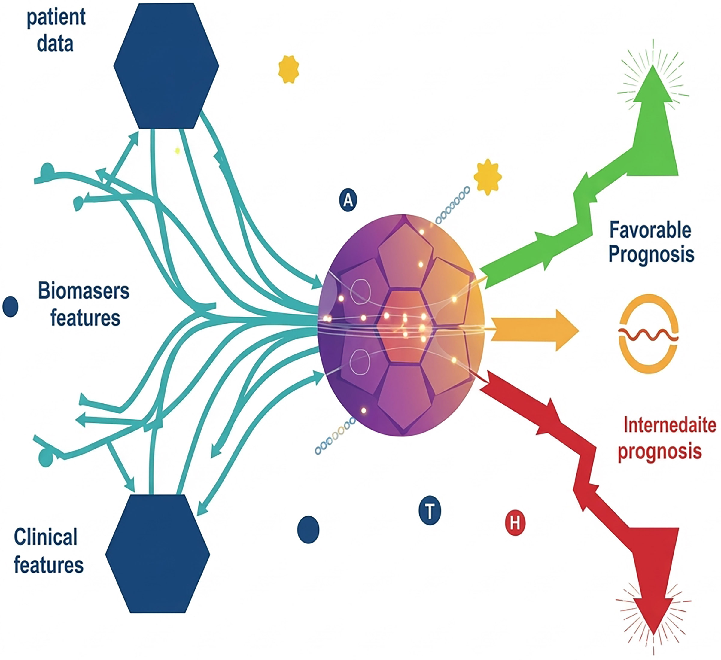
Background: Hypopharyngeal squamous cell carcinoma (HSCC) is the most frequent histologic subtype of hypopharyngeal carcinoma and has a poor prognosis. With the development of upper gastrointestinal endoscopy, an increasing number of early HSCC cases can be diagnosed and treated by endoscopic mucosal resection (EMR) and endoscopic submucosal dissection (ESD). However, clinicians lack an effective tool to predict lymph node metastasis (LNM) of early HSCC.
Methods: A total of 410 early HSCC cases were extracted from the Surveillance, Epidemiology, and End Results (SEER) database. Clinical data (50 early HSCC patients) from our hospital were applied for external validation. Univariate and multivariate logistic regression analyses to filter the risk factors were performed. A nomogram model was built to predict LNM in early HSCC patients according to the multivariate logistic regression model.
Results: HSCC patients with lymph node metastasis had worse overall survival rates and a higher risk of long-term mortality (P =0.016). The C-index and AUC of our predictive nomogram model were 0.674 and 0.623-0.726, respectively. The AUC, sensitivity, specificity, PPV, and NPV were 0.680 (95% CI: 0.525-0.835), 80.0%, 46.7%, 50.0%, and 77.8%, respectively, for the validation set.
Conclusion: Our study resulted in a novel nomogram model for predicting LNM that can be a promising tool for assessing the feasibility of EMR/ESD in early HSCC.
Impact: Our study provides a novel method for HSCC preoperative assessment before ESD.
Keywords: Metastasis; Hypopharyngeal; SEER; Nomogram; Prognosis
Introduction
Hypopharyngeal carcinoma (HPC) is a type of head and neck malignant tumour [1]. Hypopharyngeal squamous cell carcinoma (HSCC) is one of the most frequent histologic subtypes of hypopharyngeal carcinoma, accounting for up to 95% of all cases with the worst prognosis and distal survival time, with a 5-year survival rate of only 30–35% [2, 3]. Recently, lymph node metastasis (LNM) is confirmed as a crucial factor to the distal prognosis of HSCC [4]. Based on its anatomically heterogenous structure of rich lymphatics, latent HSCC cells can grow reclusively and expeditiously, resulting in distal invasion and metastasis [4]. Up to now, pathologic biopsy still is the gold standard for the diagnosis of LNM. However, several lymph nodes in certain anatomical locations including deep intra-parotid nodes, retropharyngeal nodes or nodes located just posterior to the clavicles cannot be obtained accurately [5]. Meanwhile, identification of clinical nodal stage always accompanies with lymph node dissection and a large range of surgery. Hence, it is essential to construct a noninvasive method to predict the risk of lymph node metastasis (LNM).
With the advance development of digestive endoscopic technology, the early detection of HPC has become much more possible. Moreover, the main treatment methods, such as endoscopic mucosal resection (EMR) and endoscopic submucosal dissection (ESD), are not limited only to applications in early gastric cancer and oesophageal cancer but are also suitable for HPC [6]. However, the absence of nonlymph node metastasis is an important prerequisite for choosing EMR and ESD [7].
Thus, in our study, we firstly utilized the data obtained from the Surveillance, Epidemiology and End Results (SEER) database to build a nomogram model for predicting lymph node metastasis to provide a promising tool to use before minimally invasive therapy of early HSCC. Then we used the clinical cases from our hospital for external validation and the sensitivity and specificity were 80.0% and 46.7%, respectively.
Methods
Study design and patients baseline characteristics
The clinical data were extracted from the SEER research
plus data with 17 registries and collected via SEER*Stat software (8.4.0.1; www.seer.cancer.gov) with a private ID
(account 17443-Nov2021). Informed consent was not required for the use of these registry data, as the SEER database
is publicly available and does not include any personal information [8].
The inclusion criteria for patients in our study were as follows: (1) HSCC patients aged over 18 years were diagnosed between 2004 and 2015; (2) patients were definitively confirmed by histopathology as stage Tis, T0, or T1 (the 6th American Joint Committee on Cancer stage system); (3) the primary site codes of HSCC were recorded as C12.9, C13.0, C13.1, C13.2, C13.8, or C13.9 in the light of the International Classification of Diseases for Oncology [9]; and (4) patients with whole records of baseline data and long term survival time. The patients meeting the following criteria were eligible for exclusion: (1) patients with no surgical resection; (2) the stage of lymph nodes was Nx or blank; and (3) the tumour size was blank or unknown.
For validation, we retrospectively assessed the clinicopathological data of all 50 consecutive patients with HSCC between 2017 and 2022 at the Affiliated Hospital of Medical School of Ningbo University, China. All of the patients were received surgery and lymph node dissection. All patients provided written informed consent before the surgery and all information of the participants was collected anonymously. All methods were performed in accordance with the relevant guidelines and regulations. This study was approved by the ethics committees of the institutes where the study was conducted (IRB No. KS202210028).
Variables and outcomes
According to the stage of lymph nodes, all of the patients were regrouped into N0
or N1-3 groups. Patients were classified into ≤ 60 years and > 60 years based on the age of diagnosis. Sex was
divided into male or female. The primary site of HSCC was grouped as pyriform sinus (C12.9), postcricoid region
(C13.0), aryepiglottic fold (C13.1), posterior wall of hypopharynx (C13.2), overlapping lesion of hypopharynx (C13.8),
and hypopharynx (C13.9). Grade was classified into 4 groups: well differentiated (Grade I), moderately differentiated
(Grade II), poorly differentiated (Grade III), and undifferentiated (Grade IV). The race of patients was recorded as
white, black, Asian or Pacific Islander and others. The tumour size was grouped into ≤ 10 mm and > 10 mm.
Statistical analysis
In our study, the Kaplan–Meier method was used to compare the difference in survival
times between the N0 and N1-3 groups by the Gehan-Breslow-Wilcoxon test. Univariate and multivariate logistic
regression analyses were performed to filter the risk factors for early HSCC, calculate the P value with a 95%
confidence interval (CI) and assess the independent predictors. A nomogram was built according to the results of
multivariate logistic regression, whose performance was evaluated by the C-index and receiver operating characteristic
(ROC) curve analysis. The area under the receiver operating characteristic curve (AUC), sensitivity, specificity,
positive predictive value (PPV), and negative predictive value (NPV) were computed as well. All statistical analyses
and figures were carried out via R software (version 4.0.2). P < 0.05 was deemed statistically significant.
Results
Patient demographics and clinicopathological characteristics
In total, 410 surgically resected early HSCC
patients were included in the study. Among these 410 patients, 193 cases (47.07%) were N0, and the other 217 cases
(52.93%) were N1-3. From a longitudinal perspective, N1-3 patients were more commonly younger, male, and had a worse
degree of differentiation grade. The tumour size showed a modest association with lymph node metastasis.
Table 1 demonstrates the detailed clinicopathological characteristics noted from the SEER database.
Table 1. Characteristics of early HSCC patients from the SEER database.
| Characteristic | N0 | N1-3 |
|---|---|---|
| n (%) | 193 (47.07%) | 217 (52.93%) |
| Sex | ||
| Female | 56 (13.66%) | 45 (10.98%) |
| Male | 137 (33.41%) | 172 (41.95%) |
| Age (years) | ||
| ≤60 | 60 (14.63%) | 95 (23.17%) |
| >60 | 133 (32.44%) | 122 (29.76%) |
| Race | ||
| White | 162 (39.51%) | 178 (43.41%) |
| Black | 16 (3.90%) | 23 (5.61%) |
| Asian or Pacific Islander | 12 (2.93%) | 12 (2.93%) |
| Others | 3 (0.73%) | 4 (0.98%) |
| Differentiation | ||
| Well | 19 (4.63%) | 13 (3.17%) |
| Moderate | 113 (27.56%) | 90 (21.95%) |
| Poor | 61 (14.88%) | 106 (25.85%) |
| Undifferentiated | 0 (0%) | 8 (1.95%) |
| Site | ||
| Pyriform sinus | 109 (26.59%) | 136 (33.17%) |
| Postcricoid region | 6 (1.46%) | 5 (1.22%) |
| Aryepiglottic fold | 23 (5.61%) | 14 (3.41%) |
| Posterior wall of hypopharynx | 16 (3.90%) | 17 (4.15%) |
| Overlapping lesion of hypopharynx | 0 (0%) | 2 (0.49%) |
| Hypopharynx | 39 (9.51%) | 43 (10.49%) |
| Tumour size (mm) | ||
| ≤10 | 64 (15.61%) | 61 (14.88%) |
| 10-20 | 129 (31.46%) | 156 (38.05%) |
N1-3 patients had poor distal outcome
We first compared the distal survival time between the two groups.
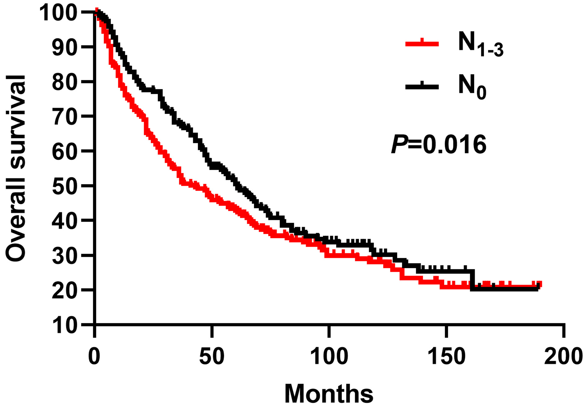
As shown in Figure 1, HSCC patients with lymph node metastasis had worse overall survival rates and
a higher risk of long-term mortality (P =0.016). The median overall survival times of N0 and N1-3 were 61 and 43
months, respectively.
Figure 1. Kaplan‒Meier analysis of LNM and overall survival time. The survival analysis displayed that N1-3 patients had poor distal outcome (P=0.016).
Risk factors related to LNM in early HSCC
To systematically predict LNM presence probability, we performed
univariate and multivariate logistic regression analyses to determine the remarkable clinical predictors based on the
clinical factors mentioned before. In the univariate model, younger age (P < 0.001), male sex (P =0.059), and
worse degrees of differentiation (P < 0.001) were associated with a higher probability of LNM in the 2004-2015 T1
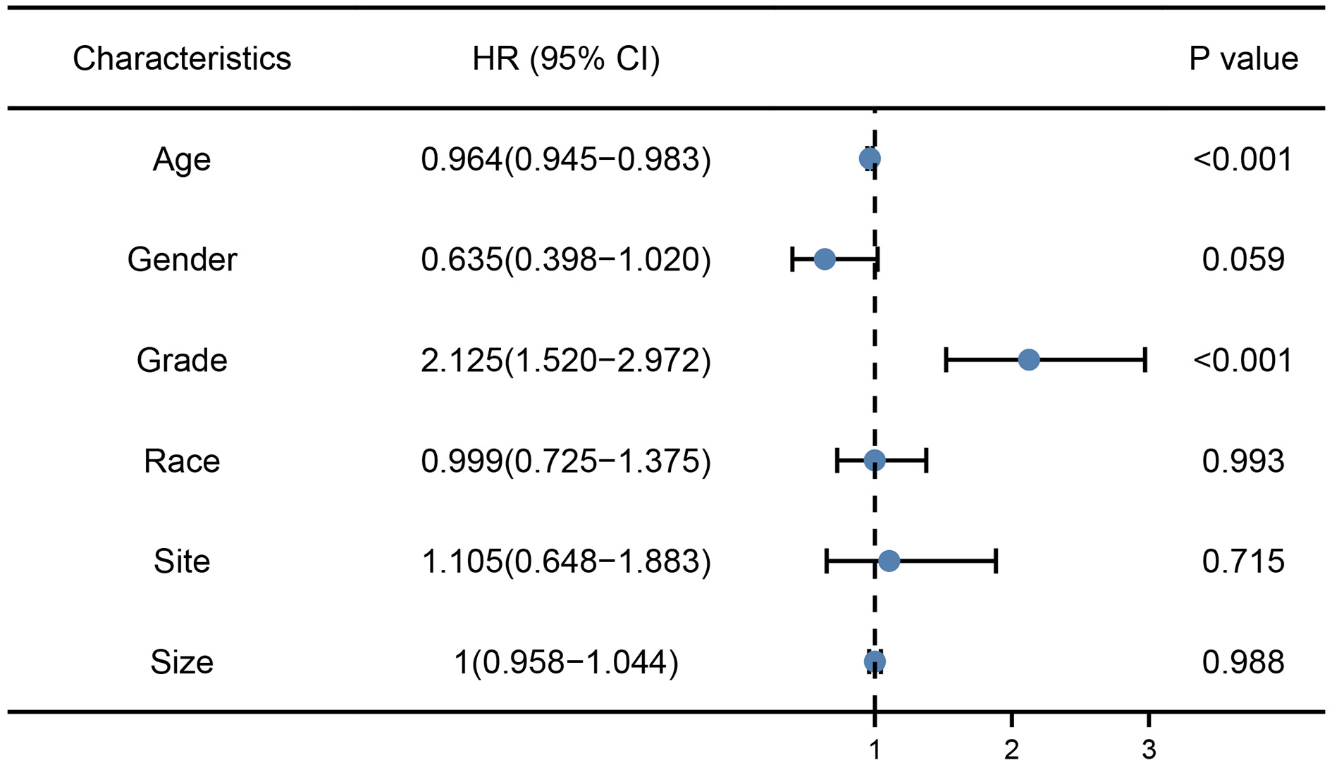
HSCC cohort (Table 2). Moreover, the results of multivariate logistic regression analysis verified
that age, sex, and degrees of differentiation were independent risk factors for assessing the probability of LNM
probability in early HSCC patients (Figure 2).
Table 2. Univariate and multivariate logistic regression analyses of factors associated with LNM in early HSCC patients.
| Variables | Univariate analysis | Multivariate analysis | ||
|---|---|---|---|---|
| OR (95% CI) | P | OR (95% CI) | P | |
| Age | 0.964 (0.945-0.963) | <0.001 | 0.964 (0.946-0.983) | <0.001 |
| Sex | 0.635 (0.396-1.017) | 0.059 | 0.637 (0.398-1.020) | |
| Grade | 2.125 (1.520-2.972) | <0.001 | 2.117 (1.531-2.929) | <0.001 |
| Site | 1.105 (0.648-1.883) | 0.715 | ||
| Size | 1.000 (0.958-1.044) | 0.988 | ||
| Race | 0.999 (0.725-1.375) | 0.993 | ||
| Constant | 0.99 | 0.998 | 3.571 | 0.100 |
Entry 0.1; Removal: 0.15, P<0.001.
Figure 2. Forest plot showing the independent clinical predictors for LNM in early HSCC patients.
Establishment and validation of the nomogram model for predicting LNM in early HSCC patients
We further
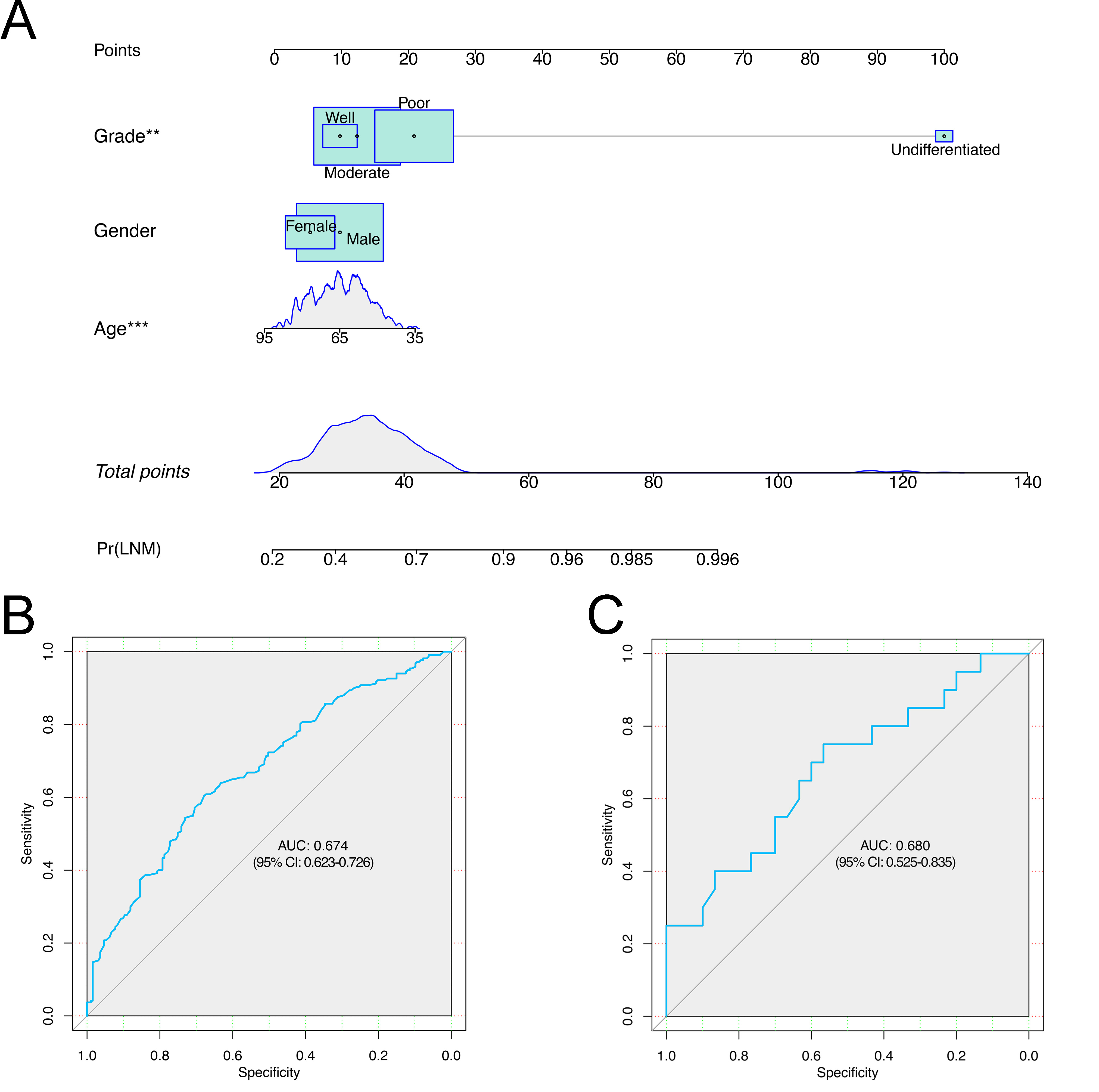
constructed a nomogram model to predict the rate of LNM in early HSCC patients according to anterior multivariate
logistic regression analysis (Figure 3A). We incorporated sex and degrees of differentiation in the
model, and clinicians could easily acquire the probability value by summing scores on each item. The C-index and AUC
of our model were 0.674 and 0.623-0.726, respectively, as displayed in Figure 3B.
To further test the actual prediction ability, we collected and used the data of our hospital as the validation sets to examine the model. A detailed description of the baseline is provided in Table 3. We drew the ROC curve, and the AUC for the validation set was 0.680 (95% CI: 0.525-0.835) in Figure 3C. The sensitivity, specificity, PPV, and NPV were 80.0%, 46.7%, 50.0%, and 77.8% for the validation set. All of the results indicated that our model had excellent adaptability to the population with early HSCC.
Table 3. Characteristics of early HSCC patients for external validation.
| Characteristic | N0 | N1-3 |
|---|---|---|
| n (%) | 27 (54.00%) | 23 (46.00%) |
| Sex | ||
| Female | 12 (24.00%) | 8 (10.98%) |
| Male | 15 (30.00%) | 15 (41.95%) |
| Age (years) | ||
| ≤60 | 12 (24.00%) | 12 (24.00%) |
| >60 | 15 (30.00%) | 11 (22.00%) |
| Differentiation | ||
| Well | 5 (10.00%) | 4 (8.00%) |
| Moderate | 13 (26.00%) | 11 (22.00%) |
| Poor | 9 (18.00%) | 6 (12.00%) |
| Undifferentiated | 0 (0%) | 2 (4.00%) |
| Site | ||
| Pyriform sinus | 10 (20.00%) | 7 (14.00%) |
| Postcricoid region | 3 (6.00%) | 4 (8.00%) |
| Aryepiglottic fold | 1 (2.00%) | 3 (6.00%) |
| Posterior wall of hypopharynx | 10 (20.00%) | 6 (12.00%) |
| Overlapping lesion of hypopharynx | 0 (0%) | 1 (2.00%) |
| Hypopharynx | 3 (6.00%) | 2 (4.00%) |
| Tumour size (mm) | ||
| ≤10 | 13 (26.00%) | 10 (28.00%) |
| 10-20 | 14 (28.00%) | 13 (26.00%) |
Figure 3. Nomogram model and ROC curves. A. A nomogram model for predicting the rates of LNM in early HSCC patients. B. ROC curve of our nomogram model. C. ROC curve of the validation set.
Discussion
The anatomic location of the hypopharynx is contiguous to the upper gastrointestinal tract, which is important for swallowing, respiration, phonation and even immunomodulation [10]. However, laryngopharyngectomy and pharyngeal reconstruction are still conventional treatment strategies, causing the loss of natural speech and dysphagia [11]. With the development of digestive endoscopy and ESD, early detection, minimally invasive surgery and preservation of anatomical function are becoming increasingly possible. Nevertheless, the absence of lymph node metastasis is the absolute indication for ESD, and it is essential to construct a satisfactory model to predict LNM [12].
In our study, we first identified that LNM significantly influenced the distal survival time for early HSCC patients. Then, a prediction nomogram model for LNM in early HSCC was built according to the SEER database. Moreover, a clinical cohort was used for external validation. The C-index of the model was 0.674 (95% CI: 0.623-0.726), and the AUC, sensitivity, specificity, PPV, and NPV of the external validation were 0.679, 80.0%, 46.7%, 50.0%, and 77.8%, respectively, which showed good discrimination and fitness. In this model, younger age at the time of diagnosis as well as worse degrees of differentiation were potentially related to a higher risk of lymph node metastasis. Younger age was related to much hypermetabolism and rich nutritional support but lower tumour doubling time, causing more aggressive biological behaviours and higher rates of LNM [13]. Degrees of differentiation and low LNM risk are the restricted condition of ESD on the basis of the Japanese Gastric Cancer Association's gastric cancer treatment guidelines [14]. All these results imply that there are many similarities in ESD between HSCC and digestive tumours.
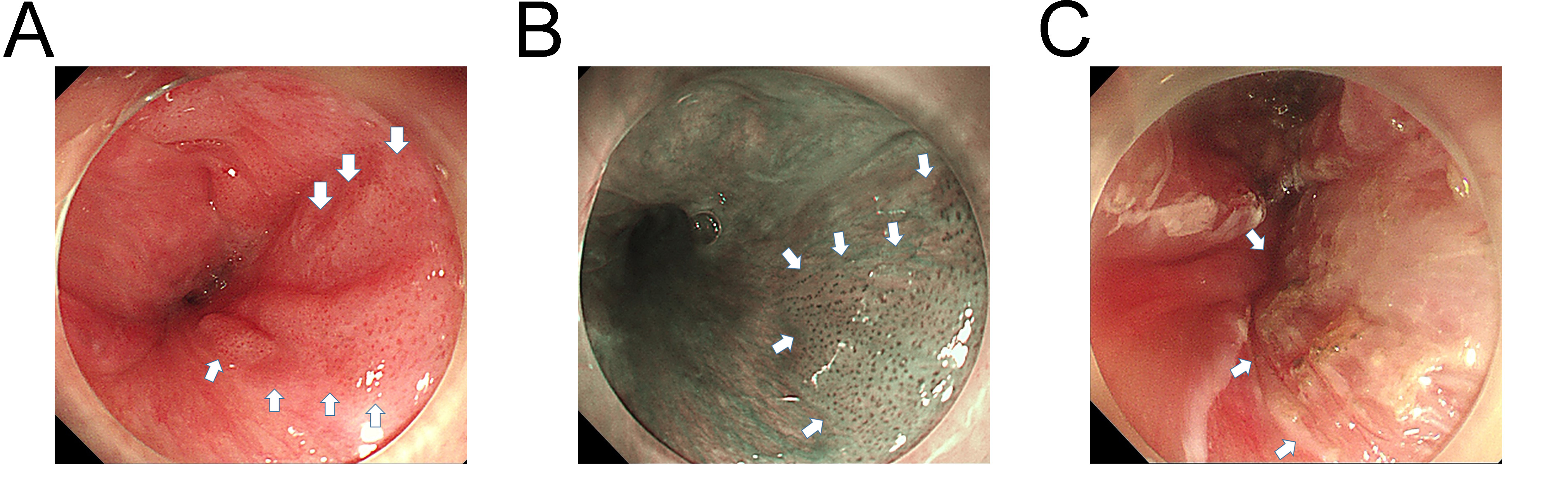
It is essential to define tumour invasion and LNM before EMR/ESD. To date, there is still no consensus among experts or clear recommendations in guidelines about EMR or ESD in early HSCC, but existing studies have clearly demonstrated their feasibility [15, 16]. At present, the otolaryngeal endoscope is still less sensitive for the detection of early or subtle mucosal and microvascular architecture changes, so it is difficult to identify the absence of early lesions. Luckily, narrow-band imaging (NBI) and magnifying endoscopy can be helpful in detecting subtle changes in the intraepithelial papillary capillary loop and brownish area in early oesophageal cancer [17]. The Japan Oesophageal Society has developed a mature method to predict the invasion depth based on magnifying endoscopic classification. JES focused on the abnormal microvessels, avascular area and intervascular background colouration, and the overall accuracy of type B microvessels in assessing tumour invasion depth was up to 90.5% [18]. This system has been widely applied in multiple centres and has high accuracy in predicting invasion depth for superficial oesophageal squamous cell carcinoma [19, 20]. More interestingly, recent studies have found that NBI can be applied in oral squamous cell carcinoma and laryngopharyngeal malignancies, which contributes to evaluating the differentiated degree and depth of invasion in early HSCC [21, 22]. Endoscopists can easily take a biopsy of the lesion in the progress of gastroscopy to obtain detailed pathological evidence, which can lay a solid foundation before EMR/ESD. Figure 4 shows the progress of diagnosing and ESD of early HSCC lesions by NBI and magnifying endoscopy using a gastroscope (GIF-Q260Z; Olympus) in our hospital. The lesion was completely resected, and the final diagnosis was carcinoma in situ (stage TisN0M0). This patient is currently under regular follow-up. In a word, we strongly recommend NBI, magnifying endoscopy and biopsy to assess and diagnose the HSCC.
Figure 4. Diagnosis and ESD of early HSCC lesions by gastroscopy. A. White light showed a 0-IIa lesion in the right pyriform sinus of the hypopharynx. B. NBI showed that the calibre of the IPCL increased notably in the lesion, which was attached to Type B1 vessels. C. The lesion was completely resected via ESD using a gastroscope.
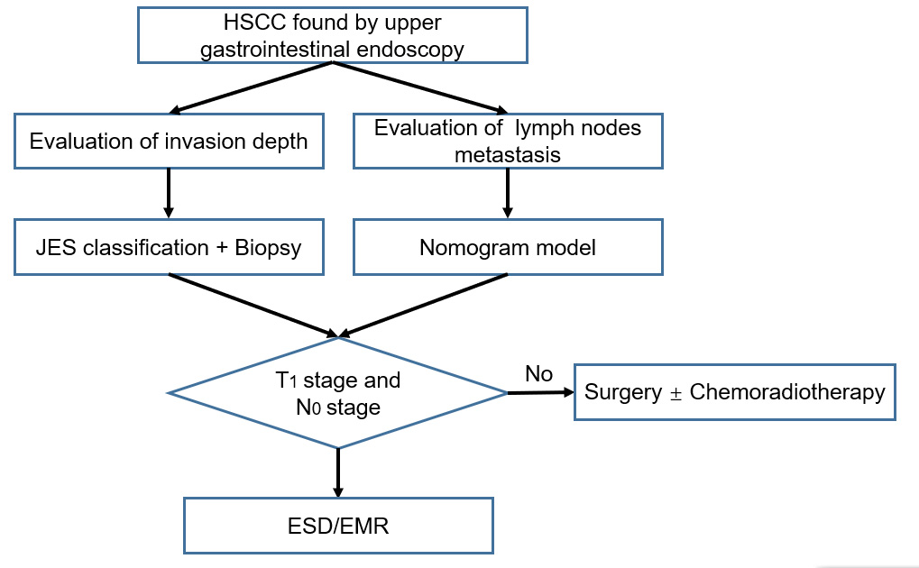
Compared to oesophageal cancer and esophagus cancer as well as gastric cancer, the depth of HPC is unrelated to the T stage, and the T number increases as the tumour size grows in cases of widespread superficial cancer on the basis of the American Joint Committee on Cancer guidelines (2017, 8th ed). The hypopharynx is prone to lymph node metastasis due to the lack of muscularis mucosa and rich lymphatic system when the tumour invades the subepithelial surface [23]. It is well reported that there is no risk of metastasis in intraepithelial cancer, and a larger HSCC correlates with a higher risk of local recurrence [24]. Moreover, on account of the narrow operation space of the hypopharynx and the requirement of ESD treatment to be performed under tracheal intubation anaesthesia, the entire hypopharynx cannot be fully exposed because of the influence of the tracheal cannula according to our clinical experiences. Hence, we selected patients with Tis, T0, or T1 stage from the SEER database for the inclusion criteria in the SEER database. The association between the depth of early HSCC and the risk of LNM should be determined in the future. As such, we propose that clinical HSCC patients are worthy to firstly receive magnifying endoscopy and biopsy to get pathological evidence. Then, our model has the capacity to confirm the indications for endoscopic treatment of early HSCC, which is worthy of validation in a larger, multicentre randomized clinical trial (Figure 5).
Figure 5. The diagnosis and treatment procedures of HSCC. We designed a novel procedure of HSCC patients received gastrointestinal endoscopy for clinical doctors.
Conclusion
In conclusion, our study resulted in a novel nomogram model for predicting LNM, which can be a promising tool for assessing the feasibility of EMR/ESD in early HSCC.
Abbreviations
AUC - Area Under the Receiver Operating Characteristic Curve; CI - Confidence Interval; EMR - Endoscopic Mucosal Resection; ESD - Endoscopic Submucosal Dissection; HPC - Hypopharyngeal Carcinoma; HSCC - Hypopharyngeal Squamous Cell Carcinoma; LNM - Lymph Node Metastasis; NBI - Narrow-Band Imaging; NPV - Negative Predictive Value; PPV - Positive Predictive Value; ROC - Receiver Operating Characteristic; SEER - Surveillance, Epidemiology, and End Results.
Declarations
Author contributions
Jianing Yan designed and performed the research and wrote the paper; Chunxia Zhu and Xueyou Lv provided the ideas about the research and supervised the report; Keshu Shan and Qier Li designed the research and contributed to the analysis of data. Keshu Shan was responsible for the revision of the manuscript for critical intellectual content; all authors issued final approval for the version to be submitted.
Acknowledgements
We sincerely thank all contributors of high-quality data to these accessible public databases.
Funding information
This study was supported by grants from the Key Scientific and Technological Projects of Ningbo [No. 2021Z133].
Ethics approval and consent to participate
This study was approved by the ethics committees of the First Affiliated Hospital of Ningbo University where the study was conducted (IRB No. KS202210028). The analysis of public data is de-identified, publicly available data from the SEER database. In accordance with US federal regulations (45 CFR Part 46.101(b)(4)), this part of research did not constitute human subjects research requiring full ethical review.
Competing Interests
The authors declare that they have no existing or potential commercial or financial relationships that could create a conflict of interest at the time of conducting this study.
Data availability
All data needed to evaluate the conclusions in the paper are present in the paper or the Supplementary Materials. Additional data related to this paper may be requested from the authors.
References
 Figures
 References
 Peer
Peer Information
Figure 1. Kaplan‒Meier analysis of LNM and overall survival time. The survival analysis displayed that N1-3 patients had poor distal outcome (P=0.016).
Figure 2. Forest plot showing the independent clinical predictors for LNM in early HSCC patients.
Figure 3. Nomogram model and ROC curves. A. A nomogram model for predicting the rates of LNM in early HSCC patients. B. ROC curve of our nomogram model. C. ROC curve of the validation set.
Figure 4. Diagnosis and ESD of early HSCC lesions by gastroscopy. A. White light showed a 0-IIa lesion in the right pyriform sinus of the hypopharynx. B. NBI showed that the calibre of the IPCL increased notably in the lesion, which was attached to Type B1 vessels. C. The lesion was completely resected via ESD using a gastroscope.
Figure 5. The diagnosis and treatment procedures of HSCC. We designed a novel procedure of HSCC patients received gastrointestinal endoscopy for clinical doctors.
Peer-review Terminology
Identity transparency: Single anonymized
Reviewer interacts with: Editor
Details
This is an open access article under the terms of the Creative Commons Attribution License(http://creativecommons.org/licenses/by/4.0/), which permits use, distribution and reproduction in any medium, provided the original work is properly cited.
Publication History
Received 2025-12-21
Accepted 2026-02-24
Published 2026-03-27